Direct inguinal hernia
Key Images
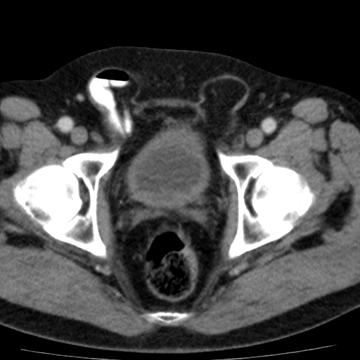
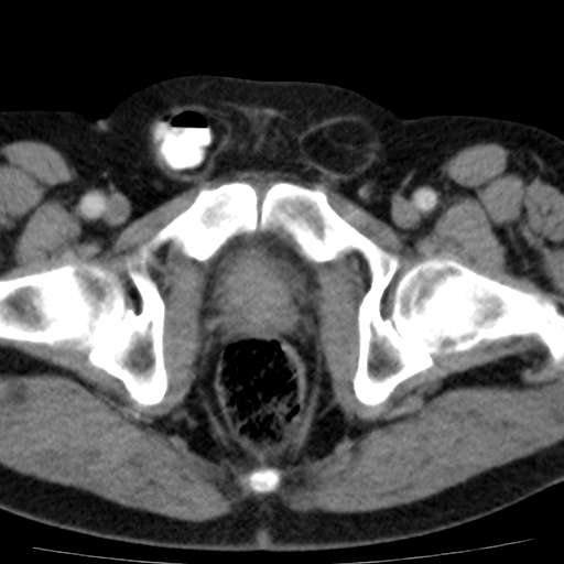
Look For First
- Peritoneal stripe deformation medial to the inferior epigastric artery on ultrasound
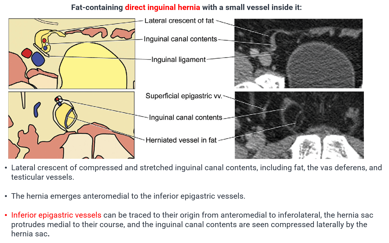
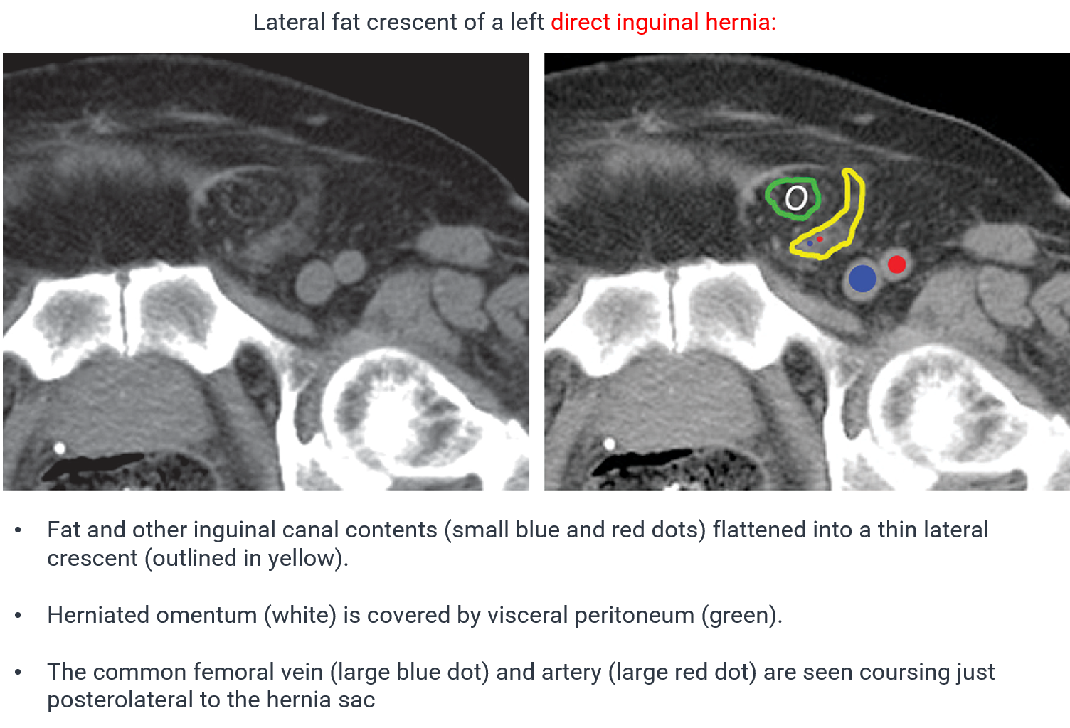
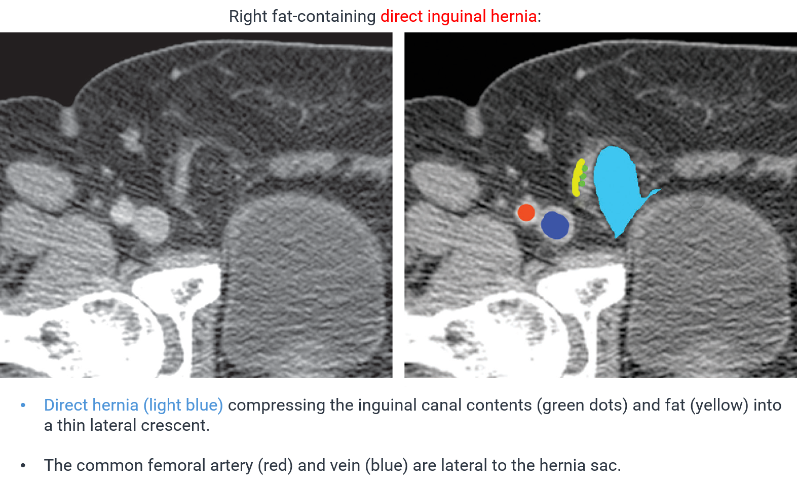
- Lateral crescent of fat sign on CT anteromedial to the inferior epigastric artery origin
- Peristaltic bowel loops visible within the hernia sac, especially during Valsalva maneuver
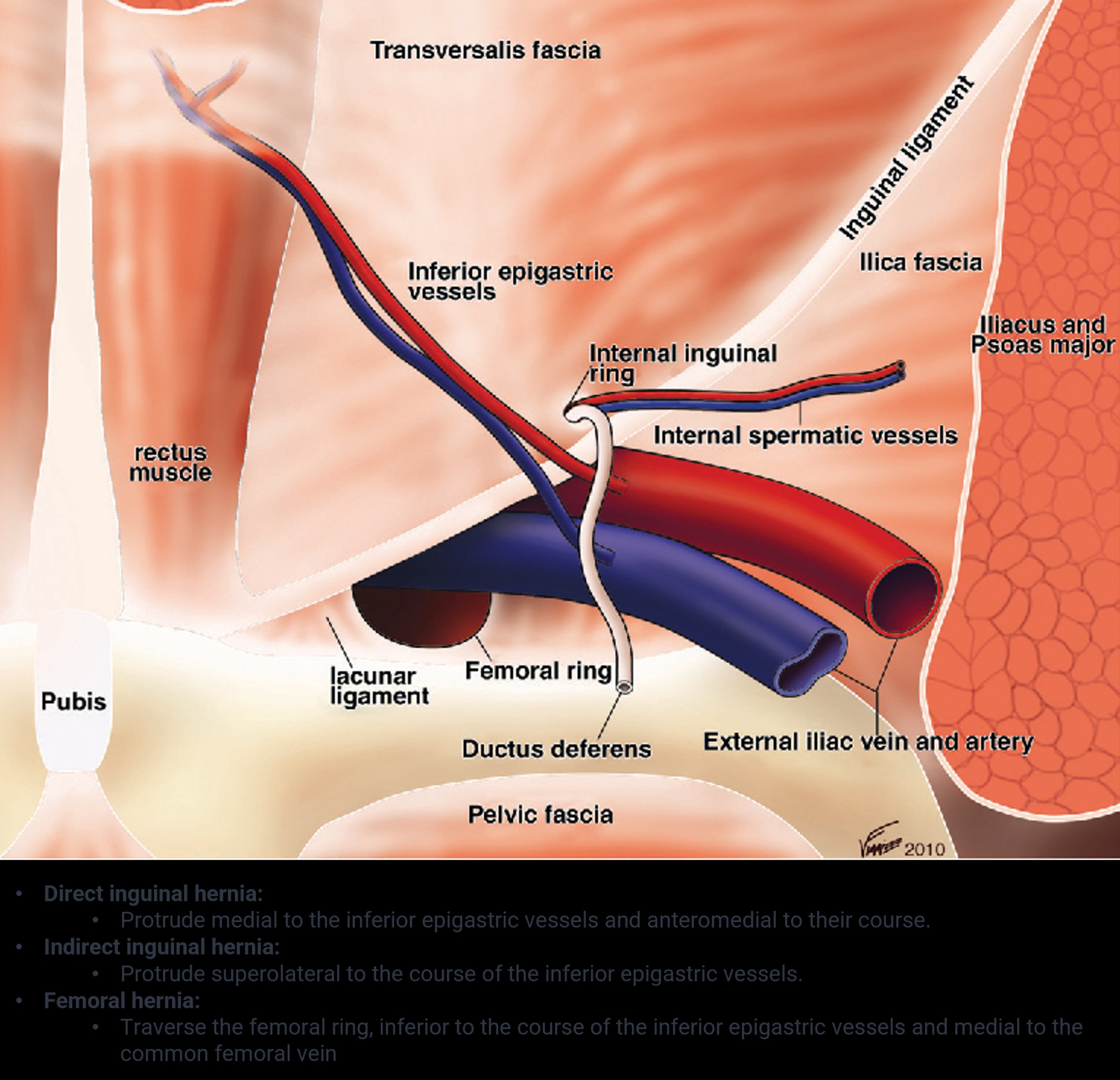
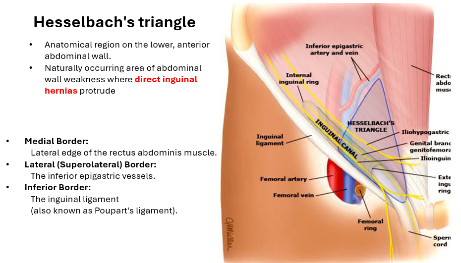
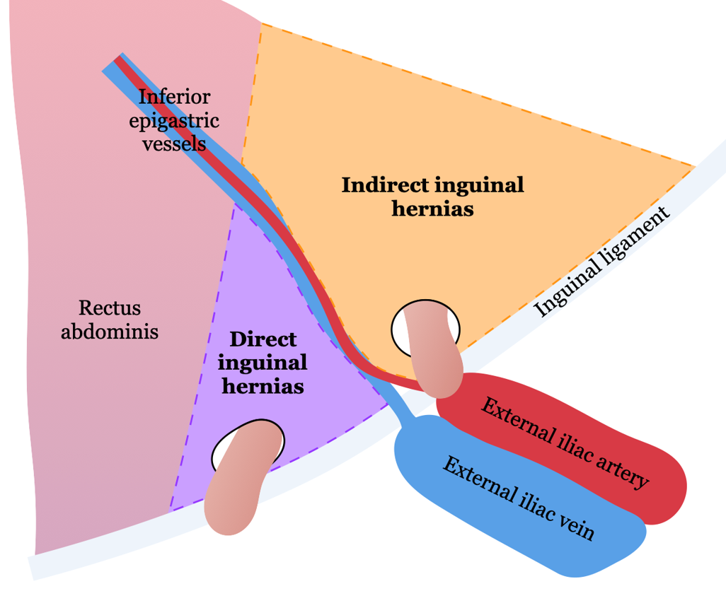
- Hernial defect in Hesselbach's triangle (medial to inferior epigastric vessels)
Key Image Findings
- On ultrasound with high-frequency linear transducers, direct inguinal hernia demonstrates deformation of the peritoneal stripe medial to the inferior epigastric artery, visible primarily with increased intra-abdominal pressure during Valsalva.
- Peristaltic bowel loops are often identified within the hernia sac as redundant, tubular structures with lamellated walls that may contain hyperechoic fat.
- The hernia sac should be interrogated for signs of incarceration with restoration of peritoneal stripe continuity following Valsalva release.
- Bowel within the hernia should demonstrate active peristalsis with detectable blood flow on power Doppler, and the bowel wall should measure <4 mm with normal layering and no surrounding free fluid.
- On CT, the lateral crescent sign—a lateral crescent of fat—is a useful diagnostic feature of direct inguinal hernia, occurring anteromedial to the origin of the inferior epigastric artery.
- Examination technique is crucial: use high-frequency transducers, perform imaging with patient supine, standing, and during Valsalva maneuver to increase sensitivity.
- The inferior epigastric artery serves as a critical sonographic landmark, identified with a cranial sweep from the external iliac artery, with the peritoneal stripe visualized just deep to this vessel.
- Direct hernias differ from indirect hernias in their anatomy: they protrude directly through the posterior inguinal wall rather than through the deep inguinal ring, so they seldom extend into the scrotum unless very large and chronic.
Differential Diagnosis
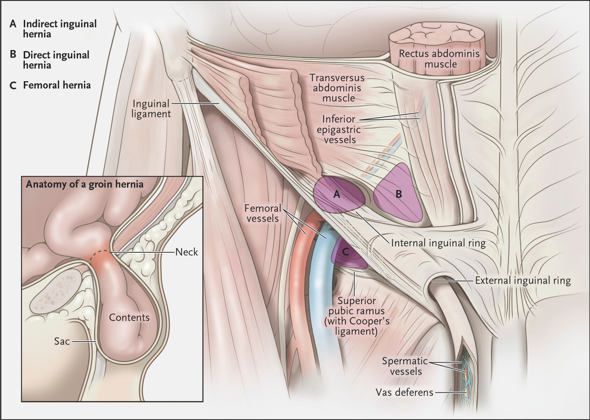
- Indirect inguinal hernia: herniates lateral and superior to the inferior epigastric vessels, does not displace the inguinal canal but rather enters it; peritoneal deformation occurs lateral to the inferior epigastric artery.
- Femoral hernia: exits below the inguinal ligament and caudal to the emergence of the inferior epigastric vessels; located in the femoral canal rather than Hesselbach's triangle.
- Busoga hernia: anatomically distinct hernia type with different location and pathology.
Discussion
Direct inguinal herniae are acquired hernias that increase in incidence with age, resulting from weakening of the transversalis fascia in Hesselbach's triangle.
They are commonly associated with chronic conditions that increase intra-abdominal pressure such as COPD, bladder outflow obstruction, and chronic constipation; increased abdominal pressure is transmitted bilaterally, so direct hernias are usually bilateral.
Compared to indirect hernias, direct hernias are less susceptible to strangulation because they often have a wide neck.
The inferior epigastric artery is the critical anatomic landmark that distinguishes direct from indirect herniae—direct hernias occur medial to this vessel.
Dynamic imaging (Valsalva maneuver) is essential for diagnosis, as the hernia may only be apparent with increased intra-abdominal pressure.
Assessment for incarceration and strangulation requires evaluation of bowel wall thickness, peristalsis, blood flow, and absence of free fluid within the hernia sac.
Reporting Pearls
Report the location relative to the inferior epigastric artery (medial vs. lateral), the size of the defect, the contents (bowel vs. fat), signs of incarceration (bowel wall thickening >4 mm, absent peristalsis, absent Doppler flow, or free fluid), and whether the hernia is reducible on dynamic imaging with release of Valsalva or probe pressure.
Pitfalls
- Failing to use proper technique: high-frequency transducers and dynamic imaging (supine, standing, Valsalva) are essential; a static supine examination may miss the hernia entirely.
- Misidentifying the inferior epigastric artery landmark, leading to incorrect localization and confusion with indirect hernia.
- Assuming a small defect with intact peristalsis and normal blood flow is safe; even apparently uncomplicated hernias require documentation of bowel wall thickness and perfusion status.
- Overlooking bilateral hernias in elderly patients with chronic intra-abdominal pressure; always examine both groins systematically.