Benign prostatic hyperplasia
Key Images
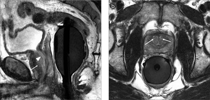
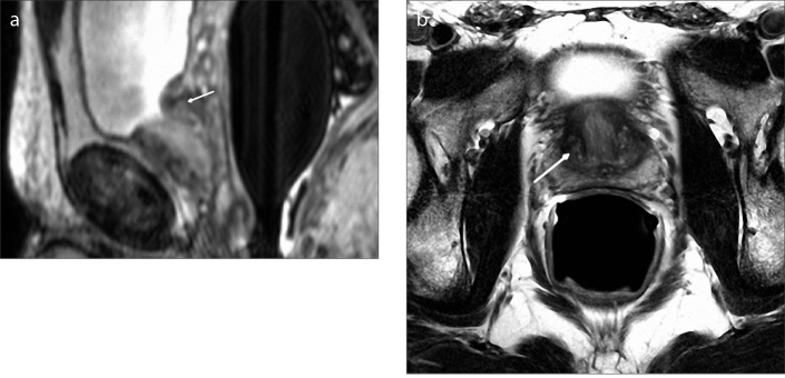
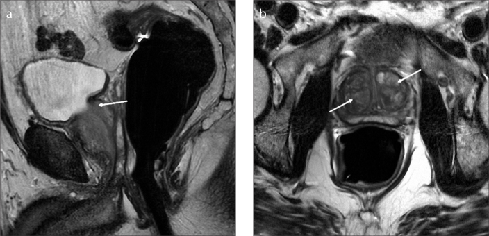
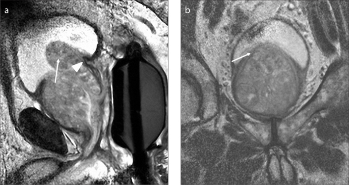
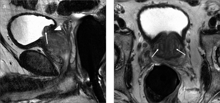
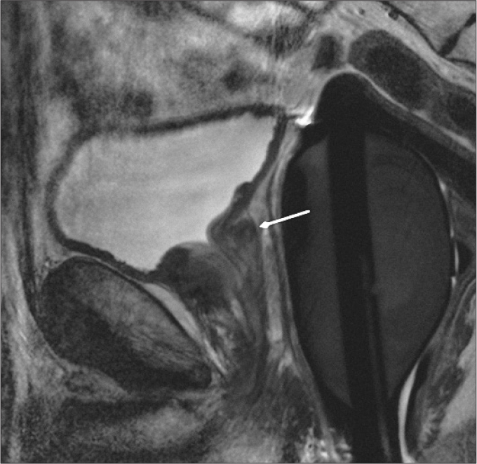
Look For First
- Identification of transition zone (TZ) versus retrourethral versus pedunculated enlargement on sagittal and axial T2-weighted images
- Hyperintense nodules (glandular/cystic components) versus hypointense nodules (stromal/fibromuscular components) on T2-weighted images
- Displacement or compression of the urethra and trigon—posterior displacement suggests TZ enlargement; superior displacement suggests retrourethral enlargement; intravesicular protrusion suggests pedunculated type
Key Image Findings
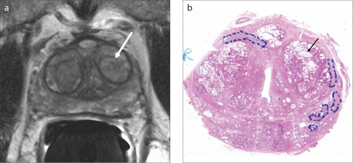
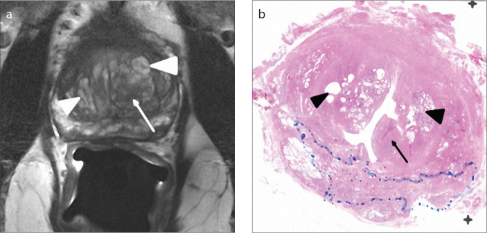
- BPH nodules appear hyperintense on T2-weighted MRI when they contain cystic ectasia and hyperplastic glandular components filled with secretion (high water content), versus hypointense when composed predominantly of fibromuscular stromal components (low water content).
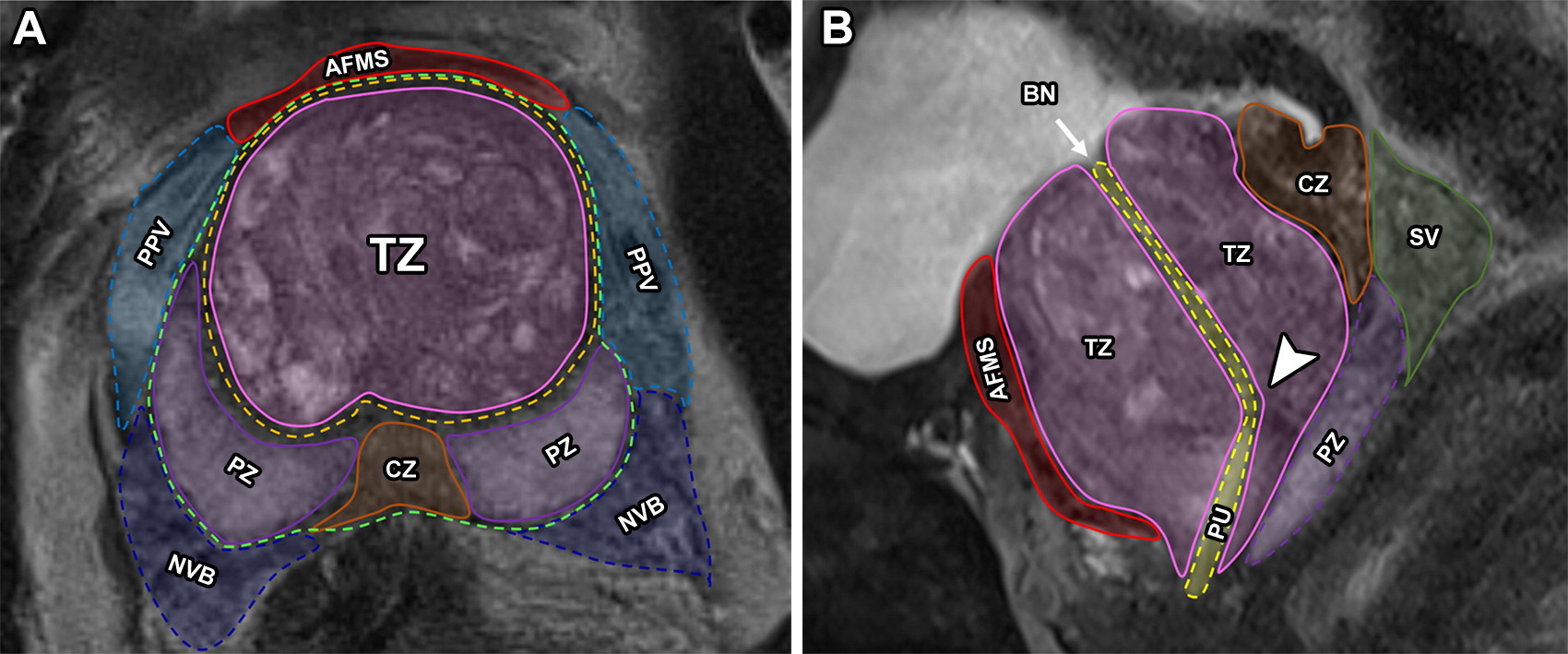
- Bilateral transition zone enlargement (Type 1, 35% of cases) shows clear TZ margins with compression of the outer muscular urethra into a fibrostromal pseudocapsule, best seen on axial and coronal T2-weighted images.
- Retrourethral enlargement (Type 2, 10% of cases) arises from deep periurethral glands posterior to the proximal urethra, displaces the trigon superiorly, and is best evaluated on sagittal images.
- Type 3 BPH (46% of cases) combines both bilateral TZ and retrourethral enlargement, representing the second most common presentation.
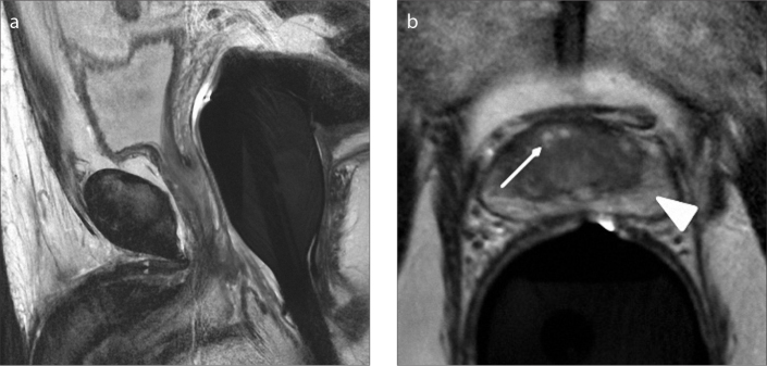
- Pedunculated enlargement (Type 4-5, more common in younger patients) presents as one or more lobulated nodules protruding directly into the bladder without superior trigon displacement, best seen on sagittal images.
- Subtrigonal or ectopic enlargement (Type 6, rare) appears as oval- or round-shaped lesion limited to subtrigonal region without inferior continuation, usually causes minimal obstructive symptoms.
- T2-weighted imaging provides superior soft tissue contrast resolution allowing differentiation of stromal versus glandular components, zonal anatomy assessment, and accurate volume estimation compared to ultrasound.
- Prostate volume classification: Type 0 shows ≤25 cm³ with little or no zonal enlargement; Types 1-7 show progressively larger volumes with varying patterns of zonal involvement.
Differential Diagnosis
- Prostate cancer versus BPH: MRI superior soft tissue contrast resolution helps differentiate by evaluating zonal anatomy and detecting suspicious nodules, though cancer can coexist with BPH.
- Stromal-predominant BPH nodule (hypointense T2) versus glandular-predominant BPH nodule (hyperintense T2): stromal nodules increase dynamic resistance while glandular nodules cause static volume effect.
- Periurethral gland (PUG) enlargement versus transition zone enlargement: PUGs located posterior to proximal urethra in superficial and deep layers; TZ located anterolateral to proximal urethra superior to verumontanum.
- Pedunculated protrusion into bladder versus retrourethral enlargement: pedunculated does not displace trigon superiorly, while retrourethral compresses and displaces trigon.
- True prostatic capsule (anatomic misnomer) versus surgical capsule: the fibromuscular pseudocapsule created by compression during BPH enlargement is not a true separable capsule despite colloquial usage.
Discussion
BPH pathophysiology involves both glandular and stromal hyperplasia: glandular enlargement causes static obstructive effect through increased prostate volume, while stromal enlargement causes dynamic effect through increased parenchymal resistance.
The MRI classification of BPH into 8 types is clinically important because different types present with different symptom profiles and require different treatment strategies, making classification essential for optimal therapy selection.
Enlargement in BPH typically begins in periurethral glands during the fourth decade, then progresses to the transition zone which becomes the main site of involvement; TZ enlargement compresses peripheral zone in most cases.
MRI allows measurement of zonal and total prostate volume, determination of stromal-to-glandular component ratio, and detection of intravesicular protrusion—all factors that guide medical versus surgical intervention.
Approximately 50% of men with histologic BPH develop lower urinary tract symptoms including both storage (urgency, frequency) and voiding (hesitancy, weak stream) symptoms affecting quality of life.
Types 1 and 3 BPH (bilateral TZ enlargement, and combined TZ plus retrourethral enlargement) are the most common presentations, accounting for approximately 81% of cases in the literature.
Reporting Pearls
Classify BPH type using systematic approach: assess transition zone enlargement (bilateral vs. absent) and retrourethral enlargement (present vs. absent) on sagittal images, then evaluate for pedunculated intravesicular protrusion and describe the stromal versus glandular composition and degree of urethral/trigonal displacement; document total prostate volume and specify which zones are involved to guide clinical management.
Pitfalls
- Failure to evaluate sagittal images: retrourethral and pedunculated enlargements are best characterized on sagittal T2-weighted images; axial-only assessment may miss crucial anatomic relationships with the trigon and urethra.
- Confusing retrourethral enlargement with pedunculated type: retrourethral enlargement displaces the trigon superiorly, while pedunculated protrusion into bladder does not displace the trigon—this distinction is critical for treatment planning.
- Assuming all T2 hypointense nodules are cancer: stromal-predominant BPH nodules are hypointense due to low water content from fibromuscular tissue; correlation with morphology (expansile nodules), enhancement pattern, and clinical context is essential.
- Neglecting to assess peripheral zone compression: BPH in the transition zone typically compresses the peripheral zone, which may affect interpretation of cancer detection and should be documented in reports.