Adenomyosis Imaging Spectrum
Key Images
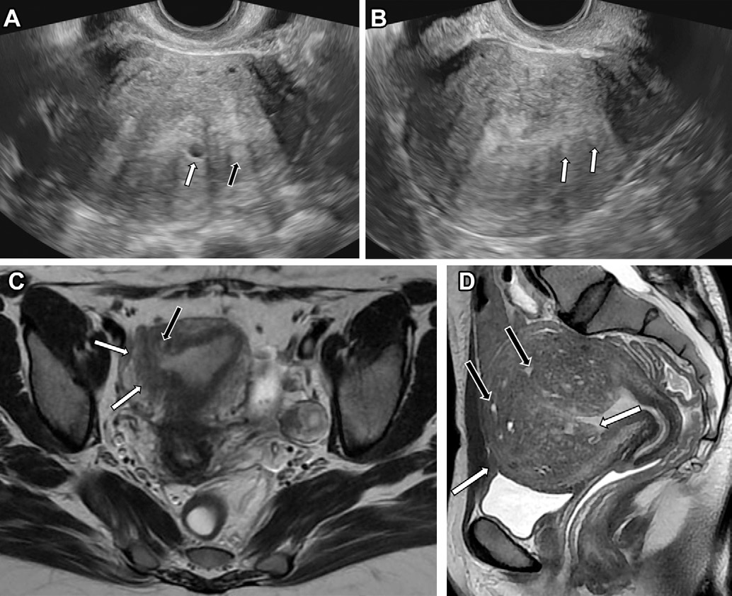
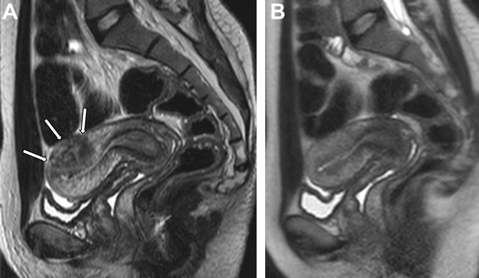
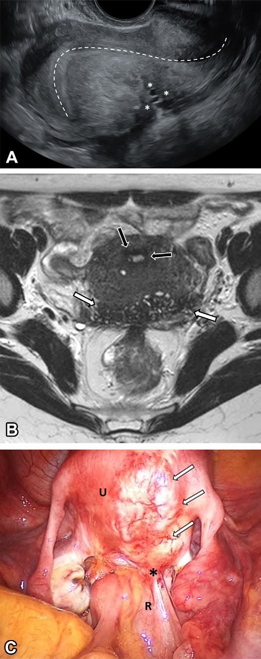
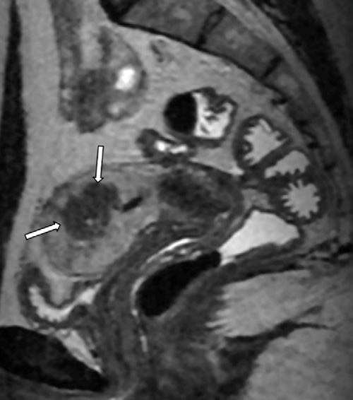
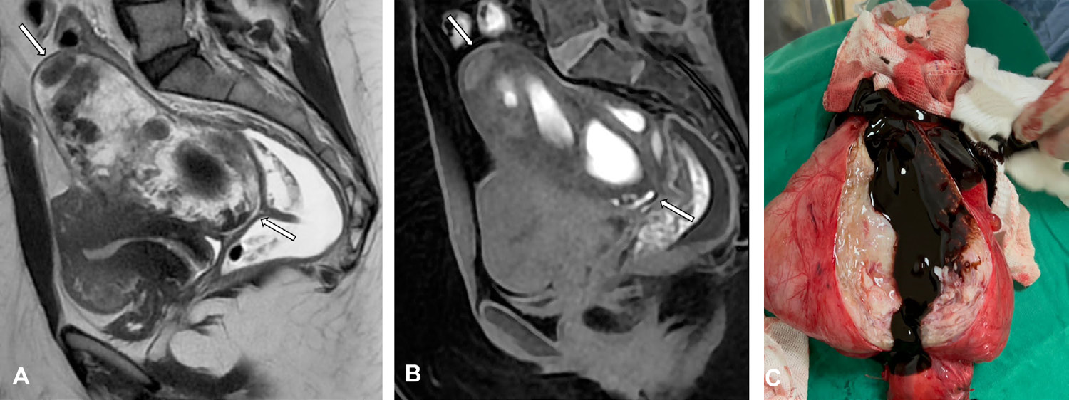
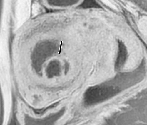
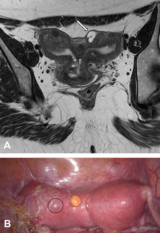
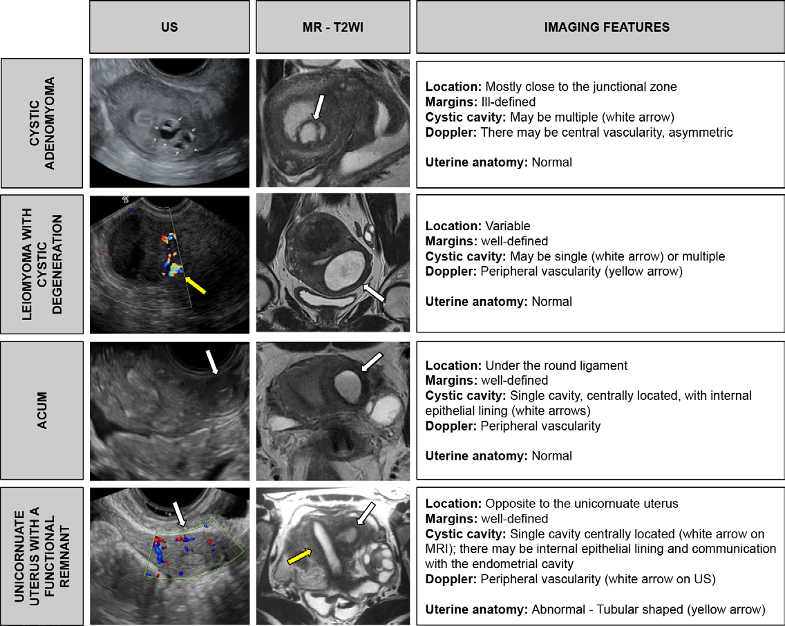
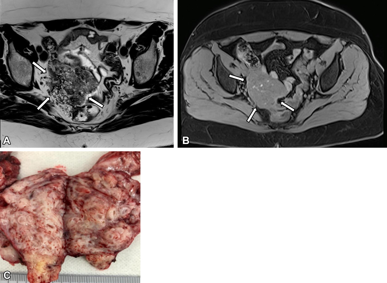
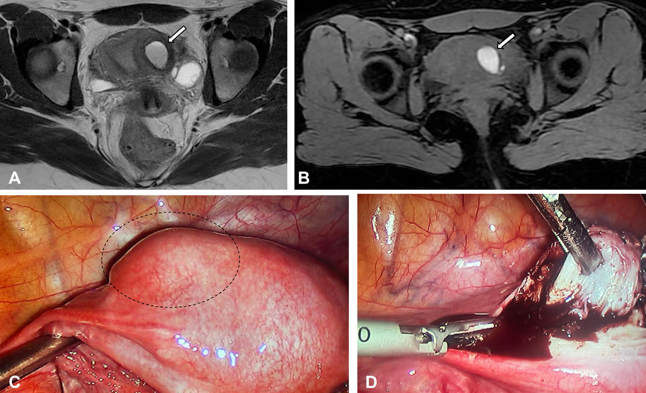
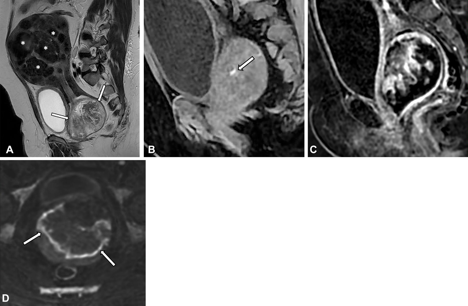
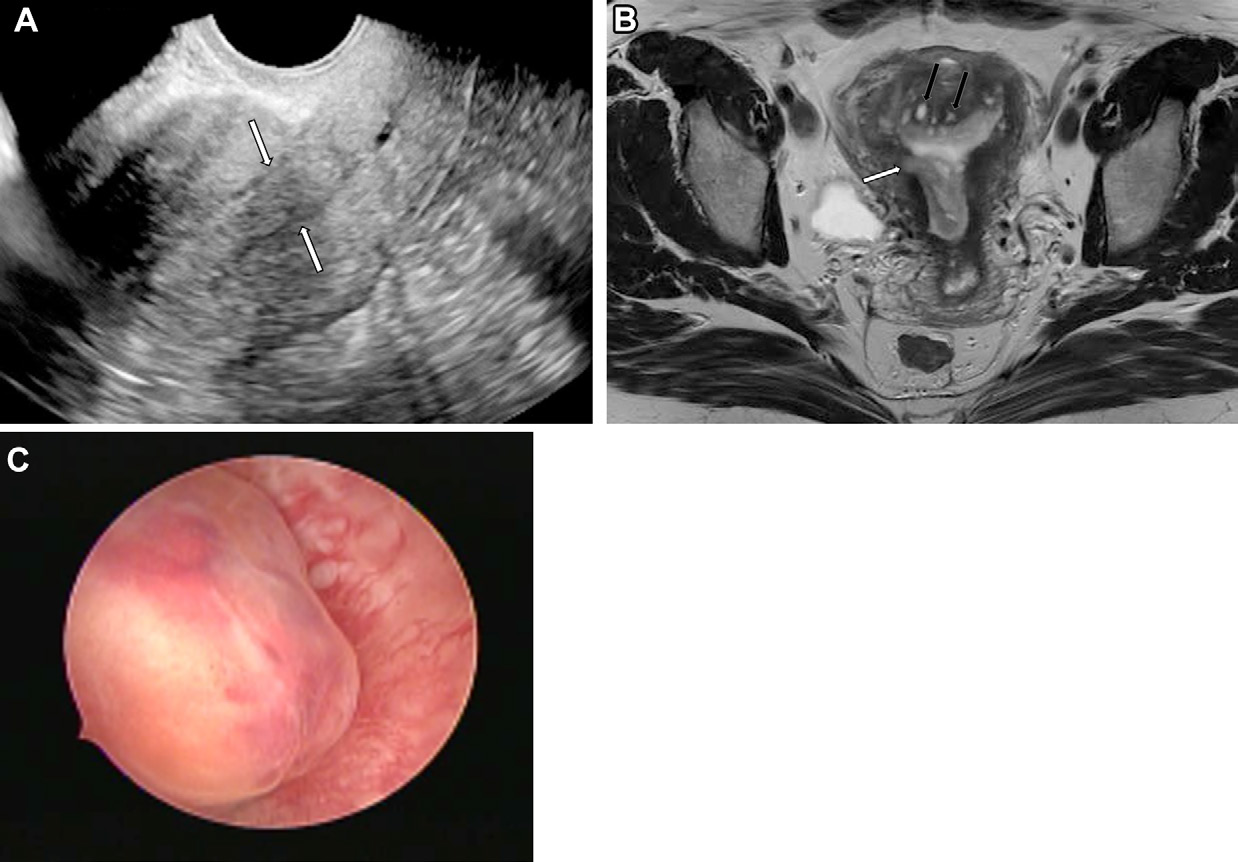
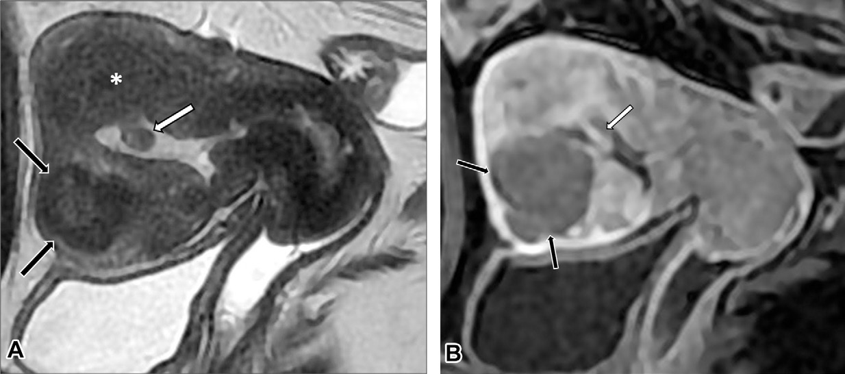
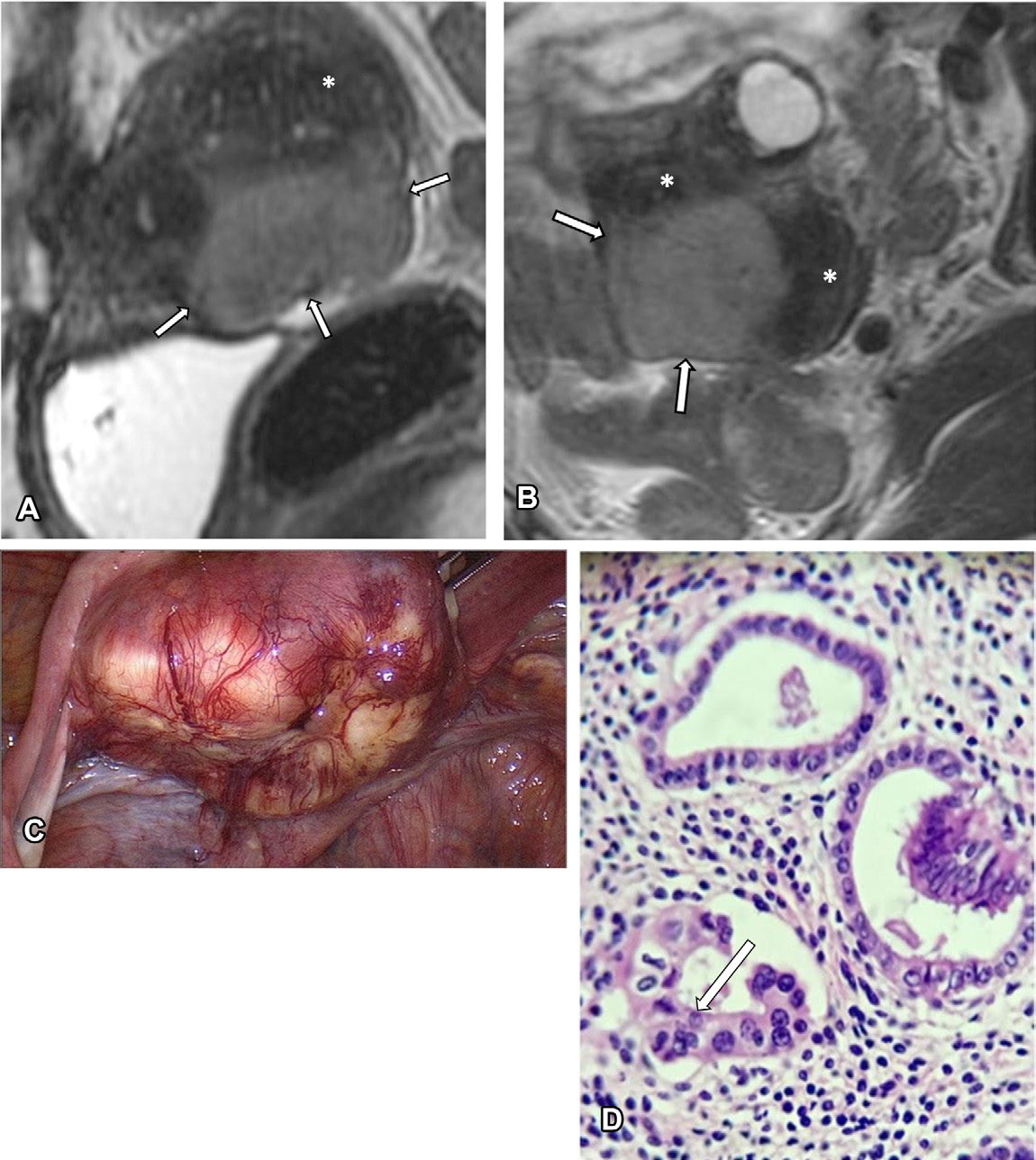
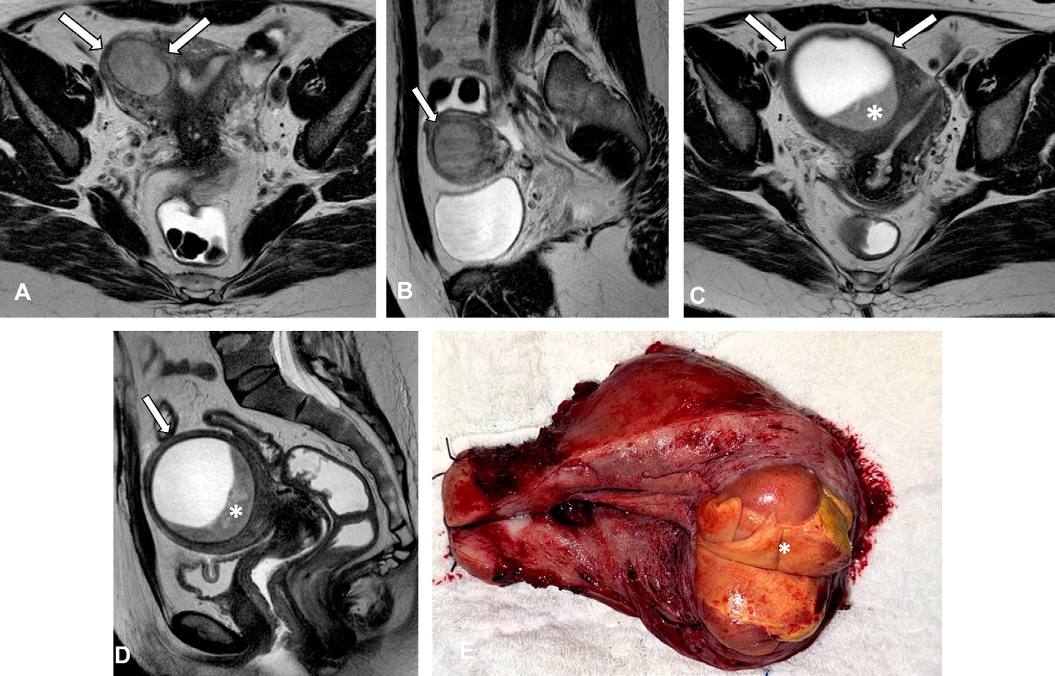
Look For First
- Junctional zone (JZ) thickening or irregularity on sagittal T2-weighted MRI or endovaginal ultrasound
- Myometrial cysts appearing as hyperintense foci on T2-weighted MRI or anechoic foci on endovaginal ultrasound
- Subendometrial hyperintense linear streaks on T2-weighted MRI or echogenic lines and buds on endovaginal ultrasound
- Ill-defined areas of low signal intensity on T2-weighted MRI or hypoechoic regions on ultrasound indicating smooth muscle hyperplasia
Key Image Findings
- On T2-weighted MRI, adenomyosis demonstrates subendometrial hyperintense linear streaks (representing ectopic endometrial glands) and myometrial hyperintense cysts (from cyclic proliferation and hormone secretion by heterotopic glands), considered the most reliable sign of adenomyosis.
- T2-weighted MRI shows ill-defined areas of low signal intensity representing smooth muscle hyperplasia and hypertrophy, often with an indistinct lower boundary between the endometrium and inner myometrium.
- Myometrial cysts frequently contain hemorrhagic content, appearing as high signal intensity on T1-weighted MRI, allowing distinction from simple cysts and supporting adenomyotic origin.
- On endovaginal ultrasound, adenomyosis presents as hyperechogenic islands, echogenic subendometrial lines and buds, myometrial cysts (anechoic foci), and fan-shaped shadowing.
- JZ thickness greater than 12 mm on MRI or indirect features including globular uterus, asymmetric myometrial wall thickening, translesional vascularity, and JZ interruption/irregularity support the diagnosis.
- Focal adenomyosis shows localized signs with greater than 25% of lesion circumference surrounded by normal myometrium and may present with JZ bulging; diffuse adenomyosis affects the entire myometrium without clear circumscription.
- MRI is superior to endovaginal ultrasound for distinguishing adenomyosis from transient uterine contractions through assessment in multiple orthogonal planes at different time points.
- Gadolinium-based contrast material aids in characterizing atypical features and distinguishing adenomyosis from leiomyomas and endometrial polyps, though not required for typical adenomyosis diagnosis.
Differential Diagnosis
- Uterine contractions—can mimic adenomyosis on single images but resolve on images obtained at different time points; MRI evaluation in multiple planes is essential to exclude this mimic.
- Leiomyomas—typically have well-defined margins, low T2 signal intensity, and may show degenerative changes, unlike adenomyosis which is ill-defined with mixed signal; gadolinium enhancement helps distinguish them.
- Endometrial polyps—appear as focal endometrial thickening without junctional zone disruption and without myometrial involvement, distinguishable by their superficial location and better-defined margin.
- Deep infiltrating endometriosis with myometrial infiltration—can cause JZ thickening and irregular margins but typically has clear endometrial origin and may have associated peritoneal endometriosis elsewhere.
- Accessory cavitated uterine masses (ACUM)—appear as cystic lesions within myometrium but lack internal epithelial lining continuity with endometrial cavity and have different clinical significance.
- Malignant transformation (sarcoma)—shows suspicious myometrial location, rapid growth, and severe adenomyotic changes; diagnosis often requires comparison with prior imaging and pathologic confirmation of adenomyotic tissue origin.
Discussion
Adenomyosis results from either invagination of basal endometrium into myometrium (leading to indistinct lower JZ boundary) or differentiation of embryonic müllerian remnants, accounting for adenomas distant from the JZ.
Risk factors include parity, miscarriages, and uterine surgical procedures (dilation and curettage, induced abortion), though the relationship with prior cesarean delivery remains debated.
Clinical correlation reveals that approximately two-thirds of patients develop symptoms (hypermenorrhea, dysmenorrhea, chronic pelvic pain) that may not correlate with disease severity, complicating clinical diagnosis.
Diffuse adenomyosis has more pronounced negative effects on assisted reproductive treatment outcomes than focal adenomyosis, emphasizing the need for standardized classification to guide management.
While histologic examination after hysterectomy remains the gold standard, endovaginal ultrasound (72% sensitivity, 81% specificity) and MRI (77% sensitivity, 89% specificity) are emerging as viable noninvasive diagnostic alternatives.
The MUSA consensus (since 2019) has established standardized terminology and classification for adenomyosis, with direct features (myometrial cysts, hyperechogenic islands, echogenic lines) being more specific than indirect features.
Reporting Pearls
When reporting adenomyosis, clearly state whether the pattern is focal or diffuse, the estimated extent of myometrial involvement (mild <25%, moderate 25-50%, severe >50%), specify the presence and location of myometrial cysts and JZ abnormalities, and note the presence of any hemorrhagic content (T1 hyperintensity) to substantiate diagnosis—this facilitates clinical correlation and guides fertility counseling.
Pitfalls
- Confusing transient uterine contractions with adenomyosis; always evaluate the JZ in multiple orthogonal planes at different time points, or use delayed imaging sequences on MRI, to resolve ambiguity between normal contractility and pathologic JZ thickening.
- Over-relying on JZ thickness alone (>12 mm) as a diagnostic criterion, which has been challenged in premenopausal women; instead, prioritize JZ irregularity and myometrial cysts as more specific markers.
- Missing adenomyosis in the cornual regions on two-dimensional ultrasound; three-dimensional ultrasound should be used when feasible to better visualize subtle JZ changes in these areas.
- Failing to administer gadolinium contrast in atypical cases; contrast-enhanced MRI is essential for characterizing unusual adenomyosis features and excluding other benign and malignant conditions such as leiomyomas and sarcoma.