Synovitis, acne, pustulosis, hyperostosis, and osteitis (SAPHO) syndrome, complicated by deep venous thrombosis
Key Images
Look For First
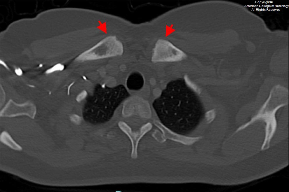
- Expanded sclerotic bone with hyperostosis at sternoclavicular junction and sternocostal joints on CT
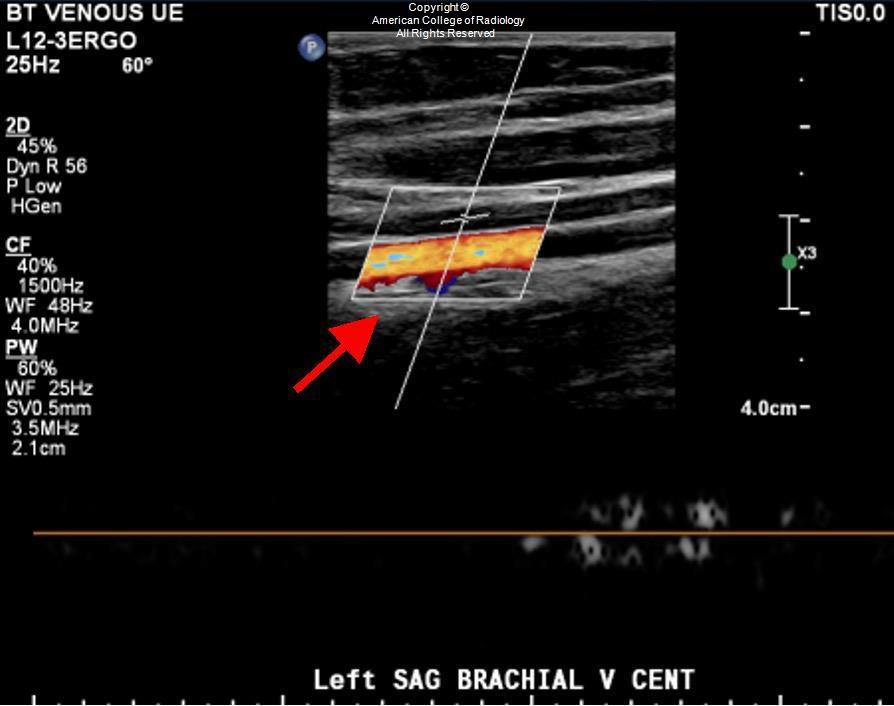
- Asymmetric or eccentric narrowing of the subclavian vein on ultrasound or CT venography
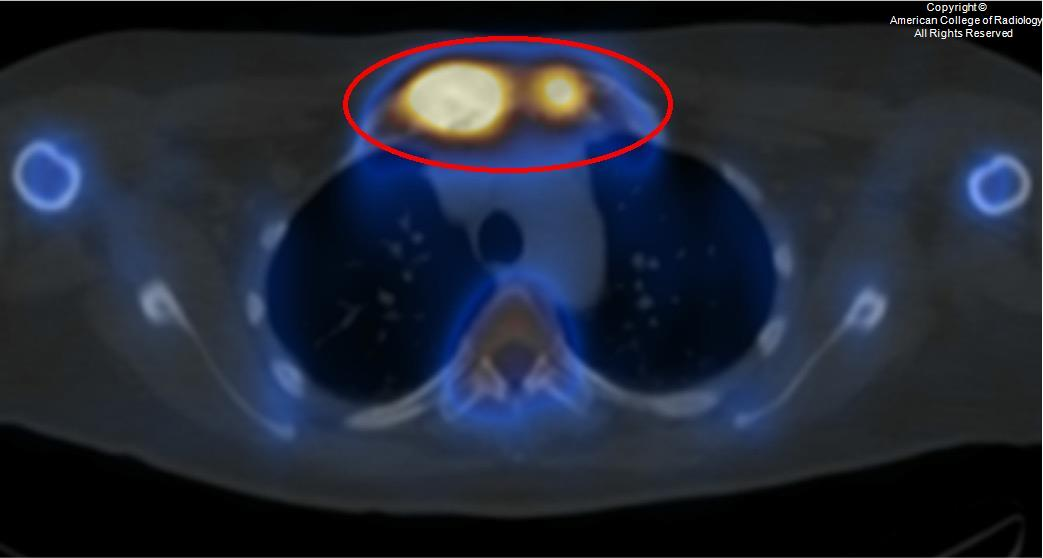
- Bull's head sign—symmetric high radionuclide uptake in bilateral sternocostoclavicular joints on bone scintigraphy
Key Image Findings
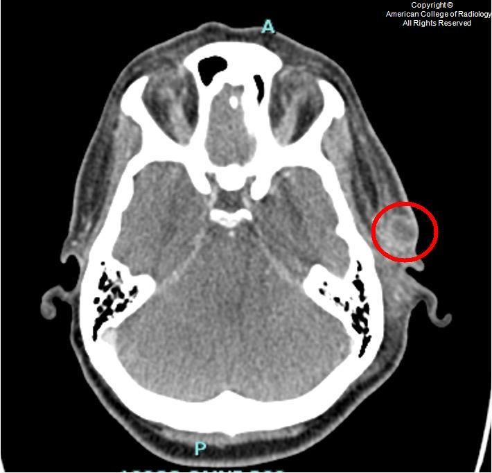
- CT shows expanded bone with sclerosis, osteolysis, periosteal reaction, and enthesopathic new bone formation at the sternoclavicular junction and first rib; sternoclavicular joint is the most frequently affected site in adults.
- Vertebral involvement includes corner cortical erosions, spondylodiscitis, and osteodestructive lesions that can cause neurological complications or spinal deformity.
- Bone scintigraphy demonstrates increased uptake in affected bone, with bull's head sign (symmetric high radionuclide uptake in sternocostoclavicular joints) considered pathognomonic for SAPHO.
- Plain radiographs have reduced sensitivity in early disease but may show polyostotic involvement; CT is the preferred method for complete evaluation of lesion extent.
- MRI detects occult lesions and soft tissue involvement; contrast-enhanced MRI improves detection of abscesses and inflammatory changes.
- Hyperostosis at sternoclavicular joint and first rib can compress the subclavian vein, precipitating upper extremity deep venous thrombosis as a rare but serious complication.
- Spine and sacroiliac joints are frequently involved after the sternoclavicular region; enthesopathy causes ligament ossification leading to bony bridging between joints.
- Axial skeleton involvement occurs in 91% of cases; peripheral joints affected in 36% of cases, with sternocostal and sternoclavicular joints most frequently involved.
Differential Diagnosis
- Osteomyelitis: lacks the characteristic sternoclavicular hyperostosis, bull's head sign on scintigraphy, and associated skin manifestations (acne and pustulosis) of SAPHO.
- Osteosarcoma: shows aggressive lytic and sclerotic changes but presents as monostotic lesion in younger patients; lacks the polyostotic involvement and systemic features of SAPHO.
- Paget's disease: demonstrates bone expansion and mixed lytic/sclerotic changes but lacks sternoclavicular predilection and systemic inflammatory features of SAPHO.
- Thoracic outlet syndrome from other causes: venous compression from hyperostosis in SAPHO is distinctive; presence of bull's head sign and skin manifestations help confirm SAPHO.
Discussion
SAPHO pathogenesis is multifactorial, involving genetic, infectious, and immunological factors; the syndrome affects any age with slightly higher prevalence in females.
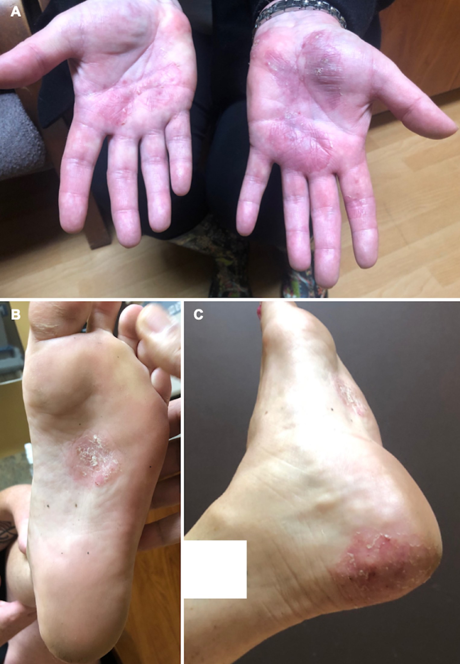
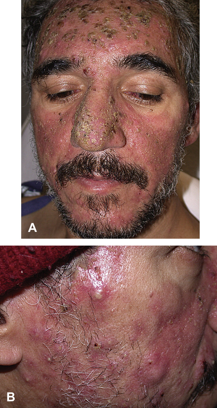
Common skin manifestations include severe palmar and plantar pustulosis and acne, which are key clinical clues accompanying osteoarticular findings.
Bull's head sign on bone scintigraphy—symmetric high radionuclide uptake in bilateral sternocostoclavicular joints—is pathognomonic for SAPHO and distinguishes it from osteomyelitis and other mimics.
Vascular complications including subclavian venous thrombosis are rare but serious; disease duration, elevated acute phase reactants, and extent of hyperostosis may contribute to thrombosis development.
Radiologists should maintain high index of suspicion for SAPHO in patients with unexplained upper extremity deep venous thrombosis, particularly when accompanied by inflammatory chest wall pain.
First-rib resection in patients with SAPHO experiencing subclavian vein compression can help prevent progressive vascular damage and is a therapeutic consideration in complicated cases.
Reporting Pearls
Report the degree and location of hyperostosis with relationship to adjacent vasculature; specifically describe any narrowing or compression of the subclavian vein, and recommend venous ultrasound or CT venography if thrombosis is suspected in the setting of sternoclavicular or first-rib hyperostosis.
Pitfalls
- Missing early disease: plain radiographs have reduced sensitivity in early SAPHO; use CT for complete assessment of lesion extent and multimodal imaging including bone scintigraphy for occult lesions.
- Overlooking thrombosis risk: deep venous thrombosis can be the initial symptom of SAPHO; maintain high suspicion when posterior triangle swelling or upper extremity symptoms accompany anterior chest wall pain.
- Confusing SAPHO with infection: osteomyelitis lacks the polyostotic distribution, characteristic sternoclavicular predilection, bull's head scintigraphic sign, and associated skin manifestations (acne and pustulosis) of SAPHO.
- Underestimating vascular compression: assess the relationship of hyperostosis to the subclavian vein on all imaging; failure to recognize compression may delay diagnosis of thrombosis and prevent timely intervention.