Amyotrophic Lateral Sclerosis
Key Images
Look For First
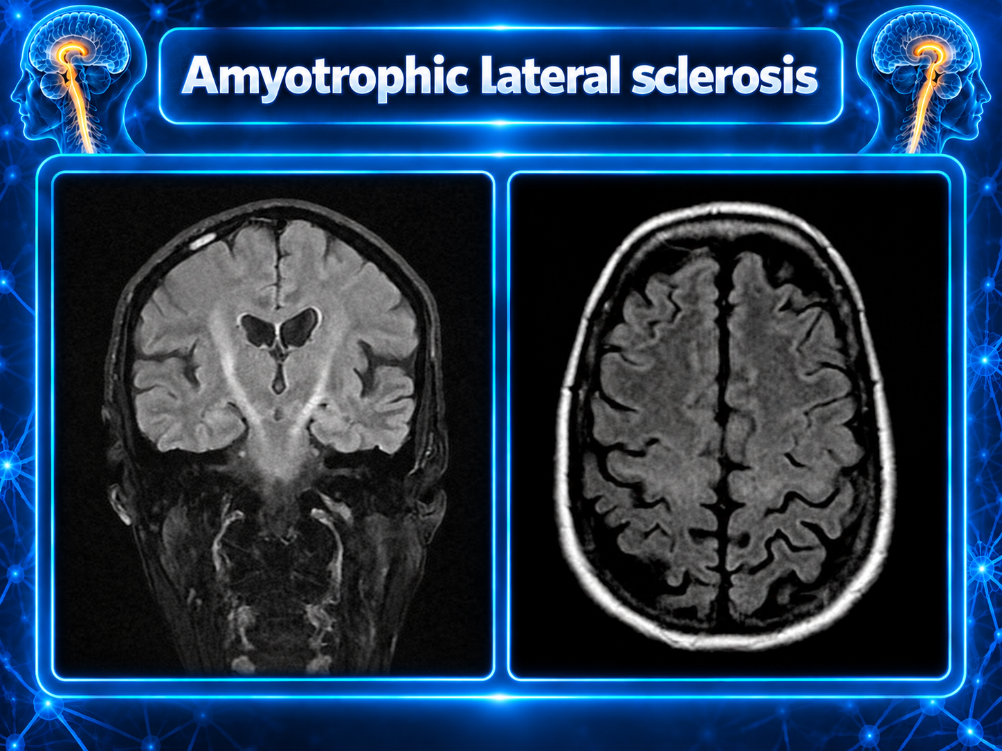
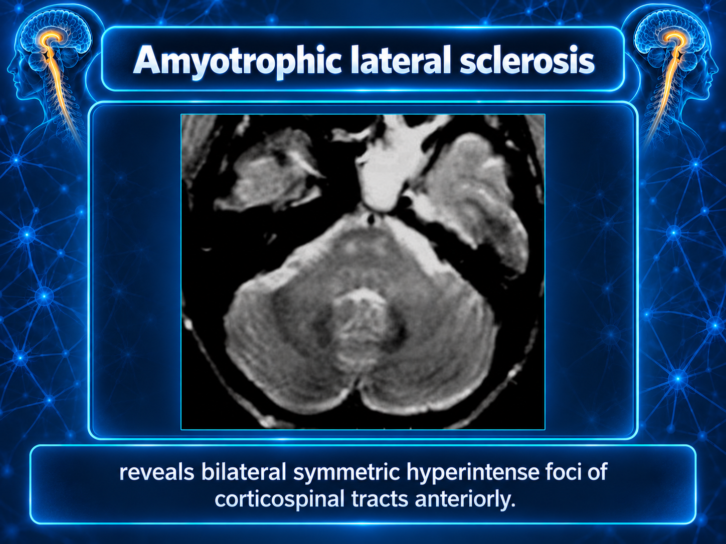
- Bilateral symmetric T2/FLAIR hyperintensities in corticospinal tracts extending from motor cortex through brainstem
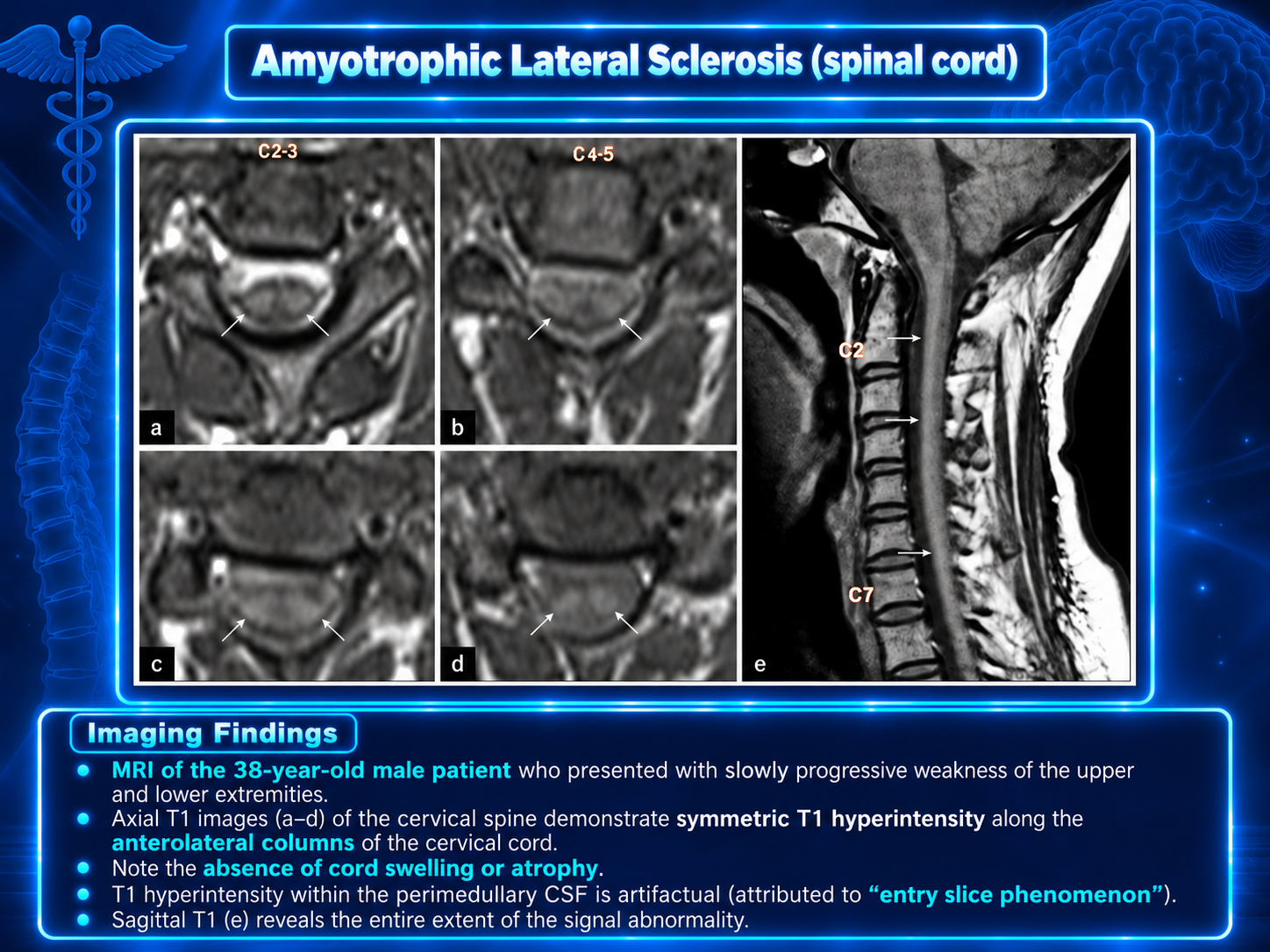
- T1 hypointensity and T2 hyperintensity in anterolateral portions of spinal cord reflecting gliosis and axonal degeneration
- Generalized decreased cerebral volume especially in gray matter of frontal and temporal lobes
Key Image Findings
- MRI
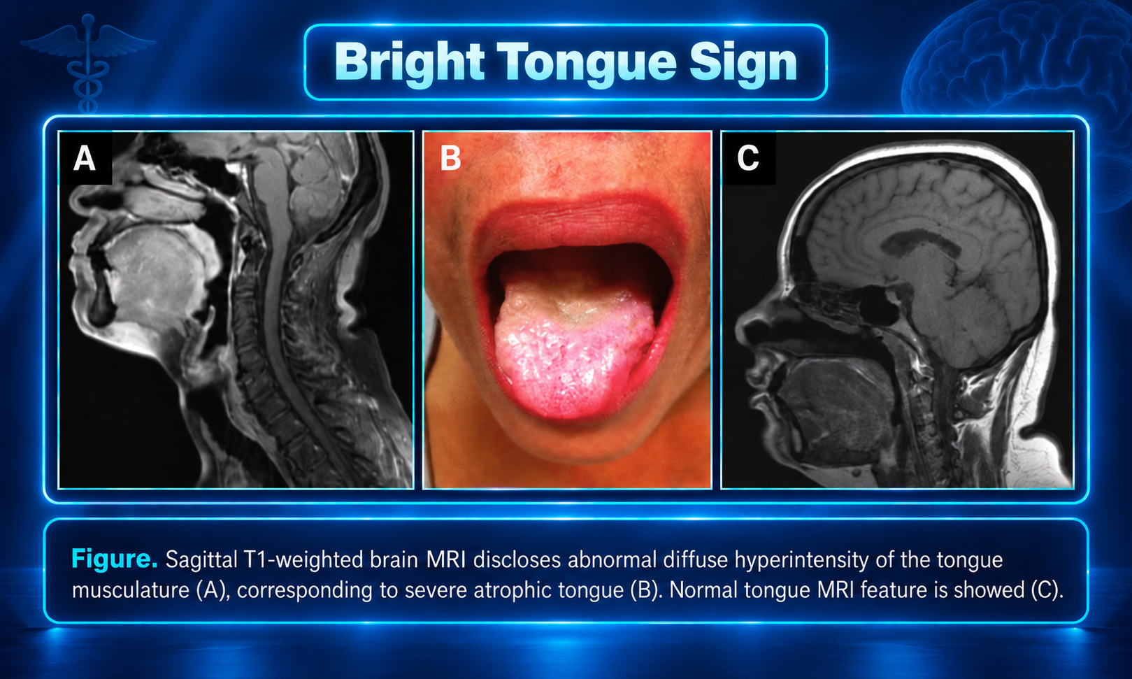
- T1: hyperintensity of the tongue may be seen in patients with bulbar involvement, known as the bright tongue sign 13
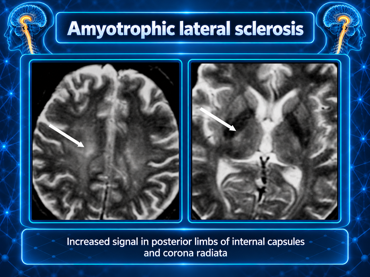
- T2: hyperintensity in the corticospinal tracts seen earliest in the internal capsule, as the fibers are most concentrated here eventually, the entire tract from motor strip to the spinal cord is affected by increased T2 signal and volume loss despite this being a well-recognized radiological feature of amyotrophic lateral sclerosis, corticospinal tract T2 hyperintensity is only seen in 30% of cases the sensitivity and specificity are rather low: specificity <70% and sensitivity <40% 6
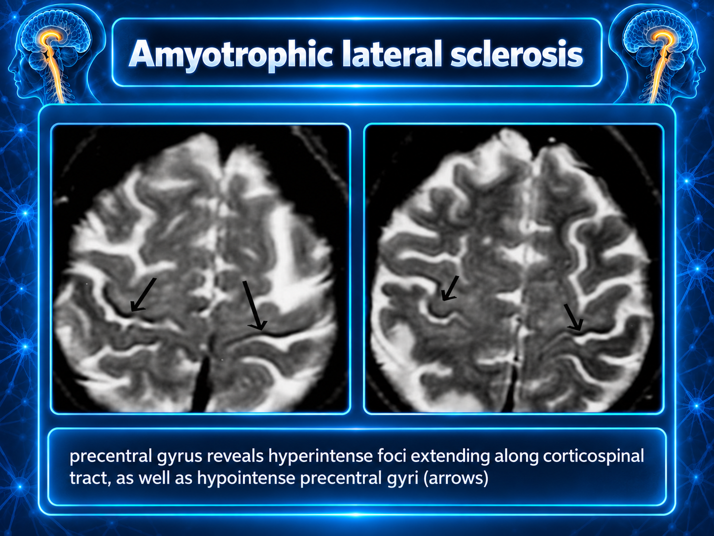
- GRE/SWI: hypointensity in the precentral gyrus bilaterally, known as the motor band sign the motor band sign may be present in patients who lack corticospinal tract T2 hyperintensity, potentially offering additional diagnostic sensitivity in this subgroup
- Generalized decreased cerebral volume has been reported, especially in the gray matter of the frontal and temporal lobes.
- Positron emission tomography (PET) has a valuable role as it demonstrates decreased radiotracer uptake within the frontal and temporal lobes in affected individuals
- Within the spinal cord, T1 hypointensity and T2 hyperintensity in the anterolateral portions are characteristic and reflect gliosis and axonal degeneration
Pathology
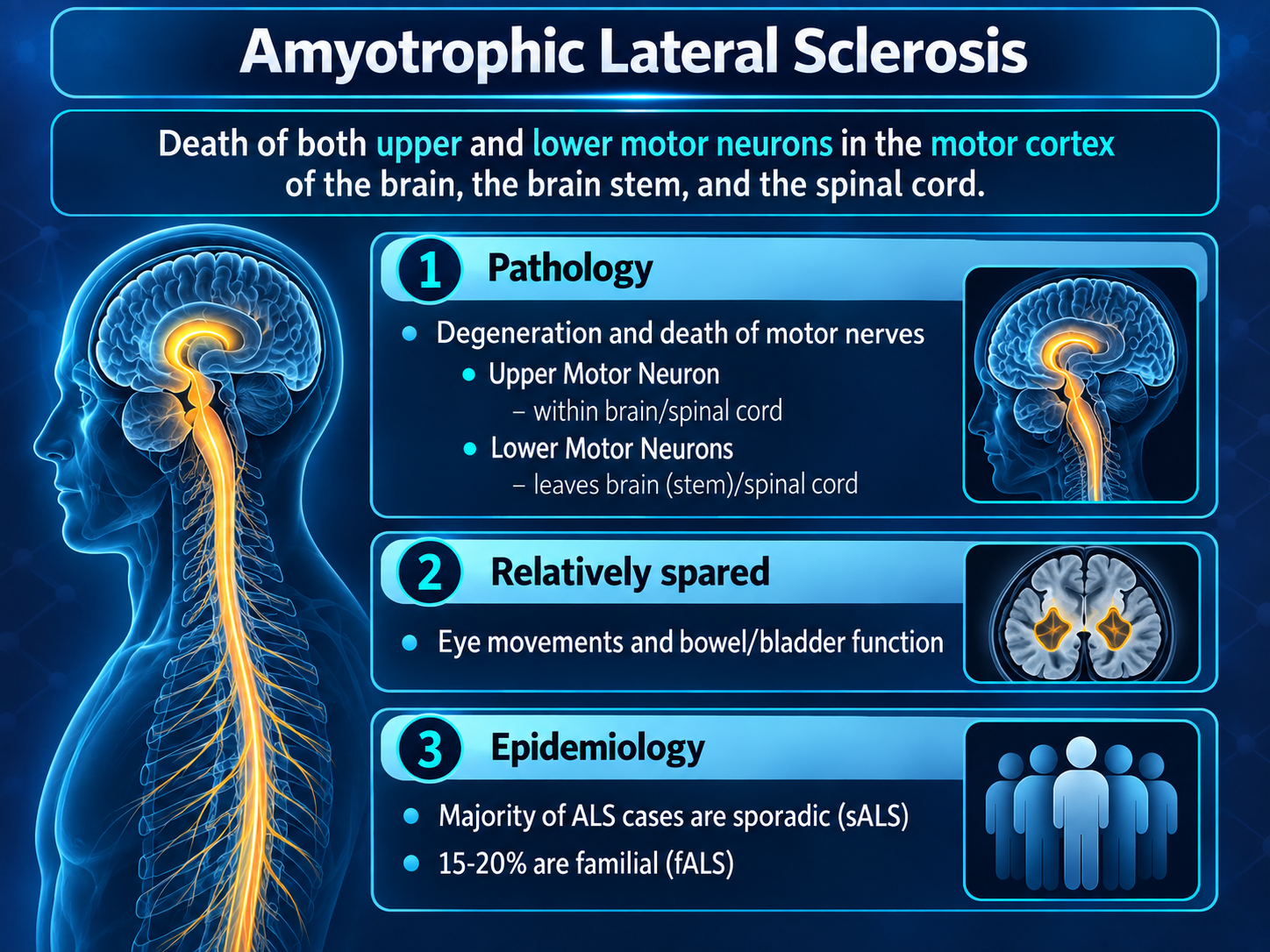
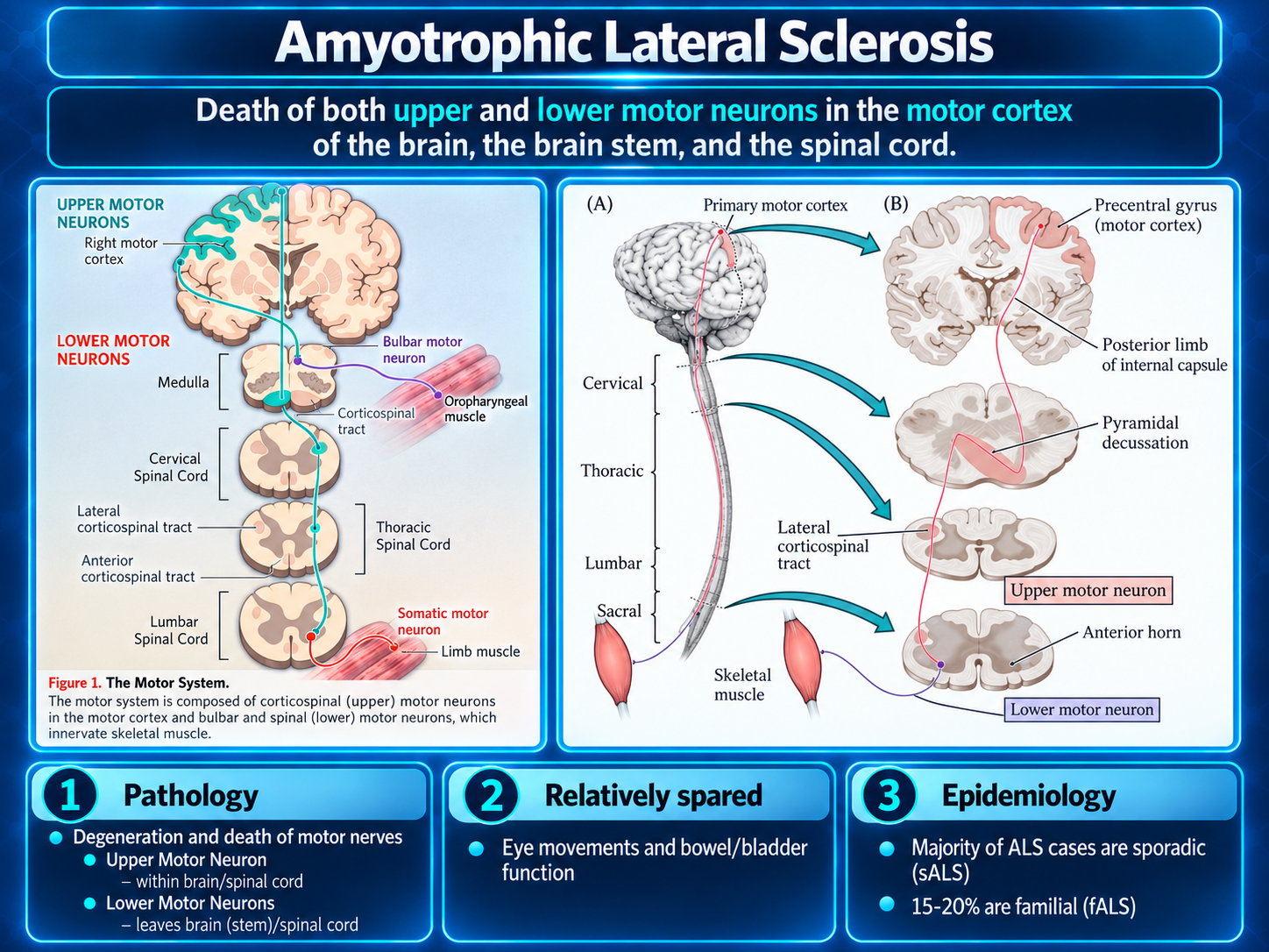
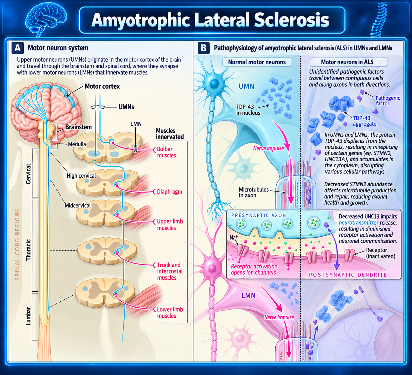
ALS is a progressive neurodegenerative disease defined by loss of both upper motor neurons (motor cortex/corticospinal tracts) and lower motor neurons (brainstem and spinal anterior horn cells). In most cases, it is also a TDP-43 proteinopathy.
Core pathology
- Degeneration and death of upper and lower motor neurons cause combined spasticity/weakness and denervation-related muscle atrophy.
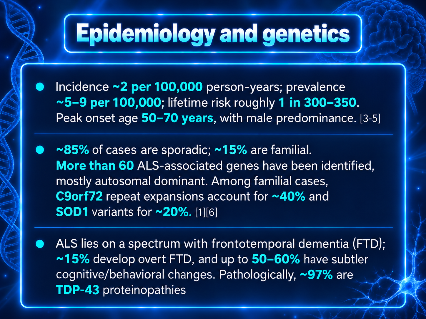
- Approximately 97% of ALS cases show abnormal loss of nuclear TDP-43 with cytoplasmic accumulation of phosphorylated, ubiquitinated TDP-43 aggregates in neurons and glia.
- The minority of cases without TDP-43 pathology typically have SOD1- or FUS-associated aggregates. C9orf72-associated ALS additionally has RNA foci and dipeptide-repeat protein inclusions.
Gross findings
- Atrophy of skeletal muscle, often affecting the tongue, bulbar/oropharyngeal muscles, and limb musculature from denervation.
- Atrophy of the motor cortex, especially the precentral gyrus.
- Pallor and sclerosis of the corticospinal and corticobulbar tracts due to axonal and myelin loss; this lateral-column sclerosis underlies the term lateral sclerosis.
- Thinning of ventral spinal roots and hypoglossal nerves, reflecting lower motor neuron loss.
Microscopic findings
- Marked loss of anterior horn α-motor neurons in the spinal cord, motor nuclei in the brainstem, and Betz cells in the primary motor cortex.
- Reactive astrocytosis and microglial activation in both gray and white matter.
- Axonal loss and myelin pallor in corticospinal tracts.
- Surviving motor neurons may contain:
- Bunina bodies: small eosinophilic, cystatin C–positive intracytoplasmic inclusions.
- Ubiquitinated/TDP-43–positive cytoplasmic inclusions.
- Skeletal muscle shows chronic neurogenic atrophy: grouped angular atrophic fibers with fiber-type grouping from repeated denervation and collateral reinnervation.
Selective neuronal vulnerability
Oculomotor neurons and Onuf’s nucleus are relatively spared until late in disease. This explains the usual early preservation of extraocular movements and bladder/sphincter function despite extensive motor neuron degeneration elsewhere.
Disease mechanisms
Although ALS has heterogeneous genetic causes, its biology converges on several interconnected processes:
- Abnormal RNA processing and TDP-43 dysfunction.
- Failure of protein quality control, ubiquitin-proteasome activity, and autophagy.
- Cytoskeletal disruption and impaired axonal transport.
- Mitochondrial dysfunction, oxidative stress, and excitotoxic vulnerability.
- Neuroinflammation, impaired nucleocytoplasmic transport, and defective DNA-damage responses.
TDP-43 pathology often follows a staged anatomic distribution, beginning in motor-system structures and extending to frontal, basal ganglia, and temporal regions as disease progresses. This pattern supports the concept of network-based, potentially prion-like propagation of abnormal protein pathology.
Differential Diagnosis
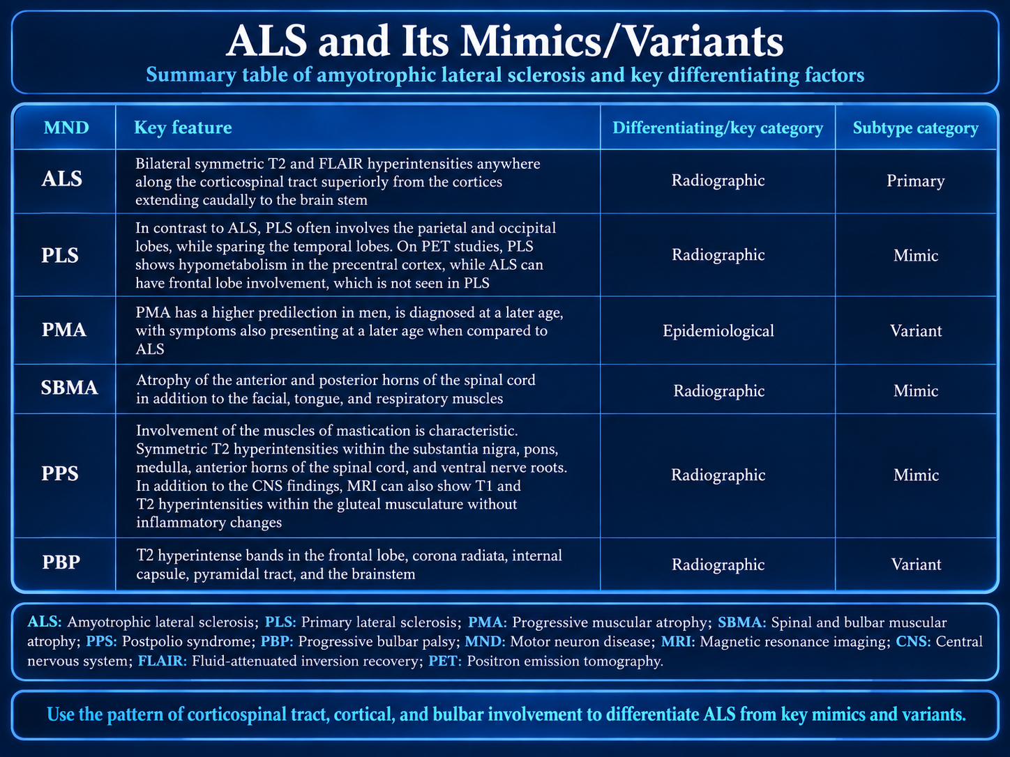
- Primary Lateral Sclerosis (PLS): shows isolated T2 hyperintensity in precentral gyrus with corticospinal tract involvement and sparing of temporal lobes, whereas ALS is more diffuse and involves temporal lobes
- Progressive Muscular Atrophy (PMA): demonstrates anterior horn T2 hyperintensities with absence of corticospinal tract involvement, unlike ALS which has bilateral corticospinal tract involvement
- Multiple sclerosis or other inflammatory conditions: conventional MRI helps exclude these conditions which mimic ALS radiographically
- Spinal and Bulbar Muscular Atrophy (SBMA): can mimic ALS clinically but has distinct genetic basis and different imaging pattern
- Postpolio syndrome: presents with lower motor neuron findings but lacks the progressive upper motor neuron involvement seen in ALS
- Progressive Bulbar Palsy: presents predominantly with bulbar symptoms but without the widespread motor neuron involvement of typical ALS
Discussion
ALS comprises 80-90% of motor neuron disease cases but has a 10% diagnostic error rate; accurate early diagnosis is critical as delayed diagnosis by average of 12 months (up to 13.1 months) leads to worse outcomes.
The pathophysiology of ALS involves prion-like protein dysregulation affecting RNA, with additional contributing factors including oxidative stress, glutamate excitotoxicity, and mitochondrial dysregulation; loss of Betz cells and CD68 macrophage aggregation are pathologic hallmarks.
Average survival is 3-5 years post-diagnosis with respiratory failure as most common cause of mortality; respiratory symptom management is mainstay of treatment.
Riluzole (sodium channel blocker) and Edaravone (antioxidant free radical scavenger) are FDA-approved agents that provide modest improvement in motor function and slow disease progression; combination therapy shows promise.
Neuroimaging plays crucial role in diagnosis by excluding other mimics and confirming ALS; conventional MRI is most useful while advanced techniques (DTI, MRS, PET) have promise but currently have low sensitivity/specificity and are not routine clinically.
Male predominance and peak incidence in sixth decade are characteristic epidemiologic features; younger patients (under 45) experience more delayed diagnosis.
Reporting Pearls
When reporting ALS on MRI, describe the bilateral symmetric T2/FLAIR hyperintensities as extending continuously along the corticospinal tracts from the motor cortex through the internal capsule, cerebral peduncle, and brainstem; note concurrent T1 hypointensity and T2 hyperintensity in anterolateral spinal cord and any associated cerebral atrophy in frontal/temporal lobes to support diagnosis.
Pitfalls
- FLAIR hyperintensity in corticospinal tracts is nonspecific and does not correlate with disease progression; do not overestimate severity based on imaging alone
- Failure to recognize that 10% of ALS cases are misdiagnosed; always correlate imaging findings with clinical presentation and exclude other MND mimics (PLS, PMA, SBMA) which have overlapping presentations
- Relying solely on conventional MRI for diagnosis when advanced techniques (DTI, MRS) have greater sensitivity; 3T DTI/MRS combination is underutilized in routine practice
- Confusing ALS imaging with dementia or other conditions affecting temporal lobes; ALS frontal-predominant changes differ from primary dementia patterns