Non-ketotic hyperglycemic hemichorea
Key Images
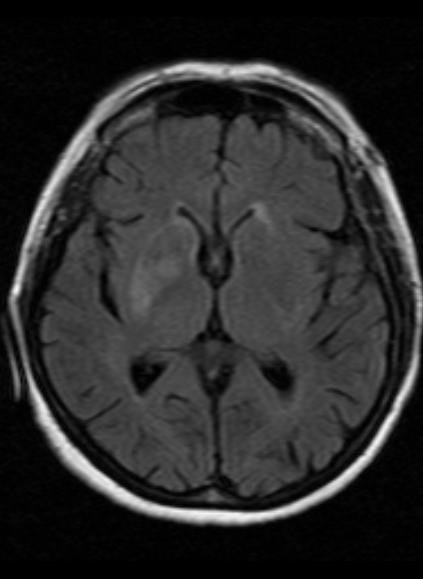
Look For First
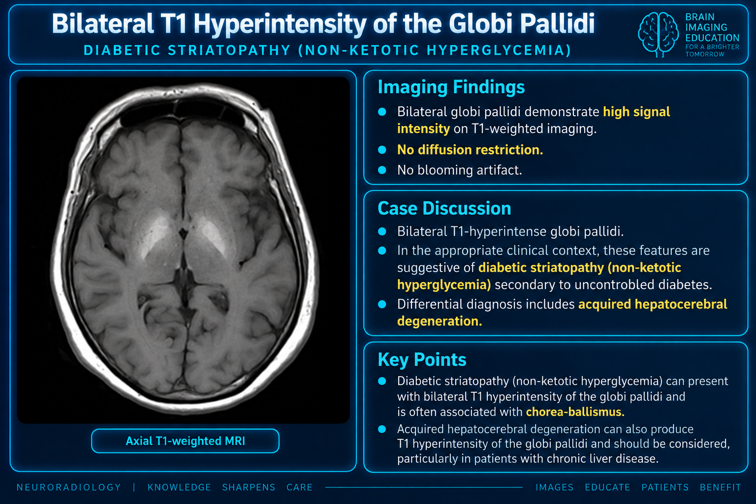
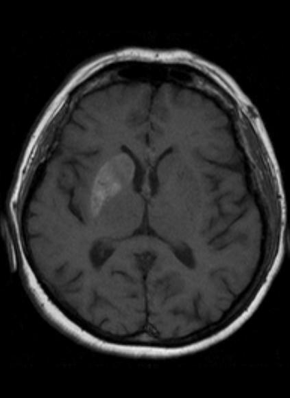
- T1 hyperintensity in the striatum (putamen > caudate) on brain MRI
- Contralateral striatal abnormality to the side of movement disorder
- Variable T2/FLAIR hypointensity in the basal ganglia despite T1 hyperintensity
- Increased susceptibility on SWI imaging in the striatum
Key Image Findings
- T1 hyperintensity is the most consistent and reliable MRI finding in non-ketotic hyperglycemic hemichorea, typically involving the putamen (95%) more commonly than caudate nucleus (40%).
- T2/FLAIR signal is variable but generally demonstrates hypointensity, creating a characteristic T1-hyperintense/T2-hypointense mismatch pattern.
- SWI imaging reveals increased susceptibility in the affected striatum, suggesting blood breakdown products or abnormal iron accumulation.
- DWI/ADC appearances are variable without a consistent pattern; restricted diffusion is not a primary feature distinguishing this condition.
- Globus pallidus involvement occurs in approximately 30% of cases and may accompany striatal changes.
- Striatal abnormalities are contralateral to the clinically affected body side in the majority of cases; bilateral changes are uncommon (~10%).
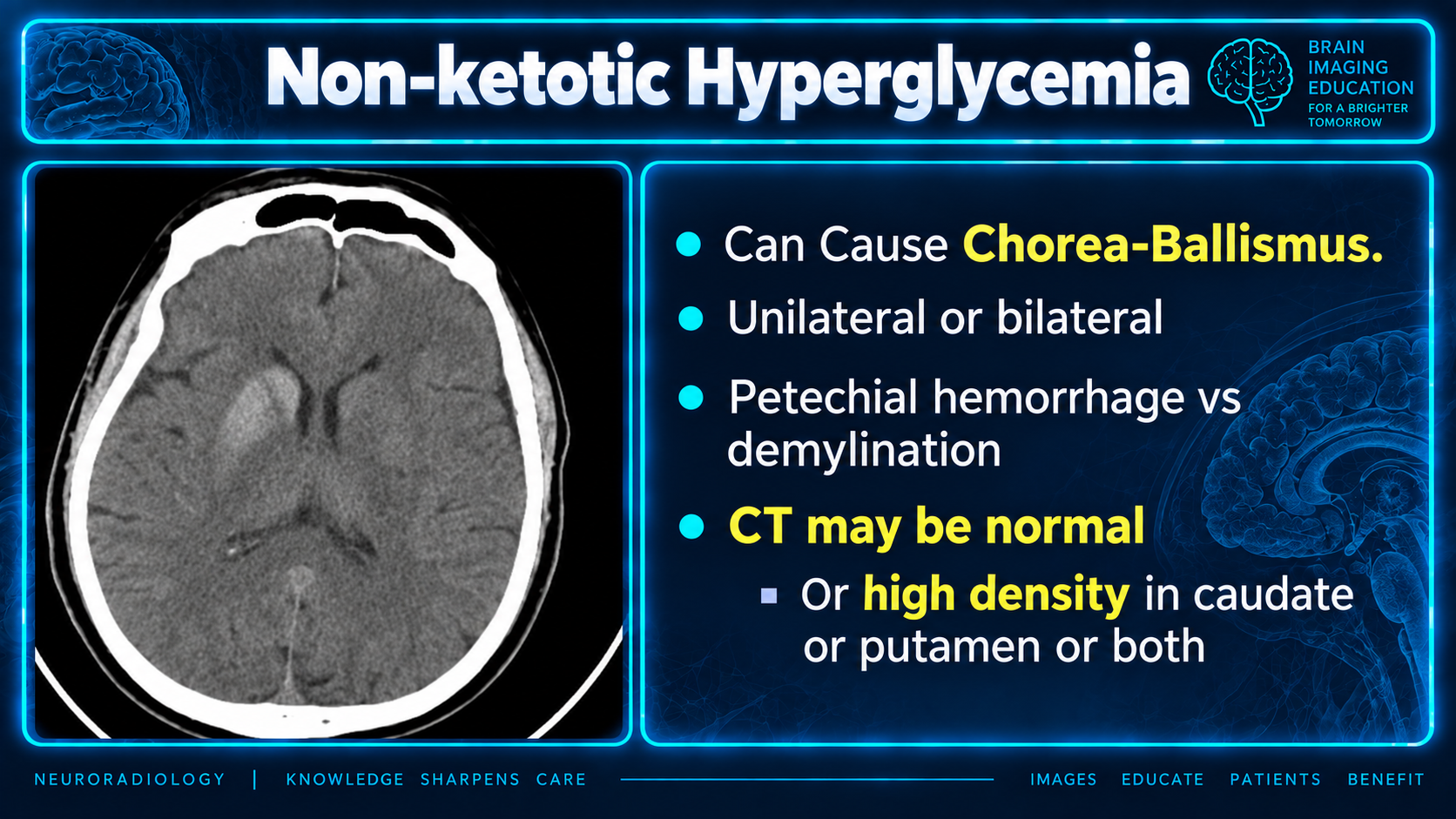
- CT brain often demonstrates striatal hyperdensity, though it is less sensitive than MRI and may initially appear normal.
- Imaging findings tend to resolve gradually with normalization of blood glucose levels, though resolution occurs more slowly than clinical symptom improvement.
Differential Diagnosis
- Wilson disease: presents with basal ganglia T1 hyperintensity but distinguished by the putamen cavitation sign, copper deposition, and associated Kayser-Fleischer rings on ophthalmologic exam.
- Striatocapsular infarct: shows acute DWI restriction and T1 hypointensity in acute phase; T1 hyperintensity develops only in chronic stages, and vascular territories do not match striatal distribution.
- Hypertensive hemorrhage: produces acute striatal hyperdensity on CT with mass effect and surrounding edema; blood products show evolution on MRI with hemosiderin on gradient echo sequences.
- Basal ganglia calcifications: appear hypointense on both T1 and T2 with calcification visible on CT; lack the T1 hyperintensity/T2 hypointensity mismatch pattern.
- Tay-Sachs disease: presents in infants and young children with progressive neurodegeneration and cherry-red macula; imaging shows widespread brain involvement not limited to striatum.
- Tuberous sclerosis: characterized by multiple cortical and subcortical tubers throughout the brain; basal ganglia involvement is less typical and associated findings aid differentiation.
Discussion
The exact pathophysiology of non-ketotic hyperglycemic hemichorea remains incompletely understood, though proposed mechanisms include hyperviscosity-induced blood-brain barrier disruption, regional metabolic damage, and altered dopaminergic receptor sensitivity.
The female predominance in reported cases may reflect augmented sensitivity of dopaminergic receptors in the postmenopausal state, linking the endocrine status to disease pathogenesis.
Neuroimaging is abnormal in approximately 93% of symptomatic patients, making MRI a highly sensitive tool for confirming the diagnosis when clinical presentation is suggestive.
The term 'diabetic striatopathy' is nonspecific and should be qualified as symptomatic (clinical + imaging), clinically isolated (clinical only), or radiologically isolated (imaging only) to clarify the clinical-radiological correlation.
Clinical symptoms typically resolve rapidly with glucose normalization and symptomatic management, whereas imaging abnormalities tend to persist longer and may take weeks to months for complete resolution.
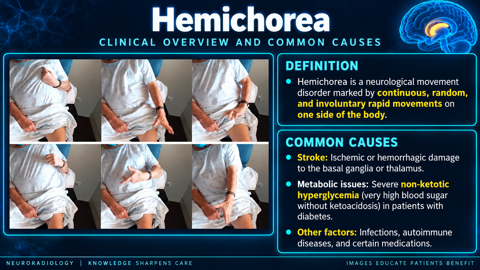
Non-ketotic hyperglycemic hemichorea is a rare neurological complication that should be suspected in elderly diabetic patients presenting with acute hemichorea, particularly in Asian populations.
Reporting Pearls
Describe the finding as "T1 hyperintensity with T2/FLAIR hypointensity and increased SWI susceptibility in the striatum (predominantly putamen), contralateral to the clinically affected limbs, in the setting of non-ketotic hyperglycemia," and emphasize that this mismatch pattern is characteristic of the condition rather than acute infarction or hemorrhage.
Pitfalls
- Misinterpreting T1 hyperintensity in the basal ganglia as Wilson disease without checking for putamen cavitation sign, copper deposition pattern, and systemic features.
- Confusing non-ketotic hyperglycemic hemichorea with acute striatocapsular infarction by overrelying on DWI; remember that DWI findings are variable in NHH and the T1 hyperintensity pattern differs from typical acute infarction.
- Overlooking the T2/FLAIR hypointensity component; the T1-hyperintense/T2-hypointense mismatch is characteristic and should prompt consideration of this diagnosis rather than other causes of striatal T1 hyperintensity.
- Assuming normal CT excludes the diagnosis; early CT may be normal, and MRI is more sensitive for detecting the abnormalities in non-ketotic hyperglycemic hemichorea.