Posterior cortical atrophy
Key Images
Look For First
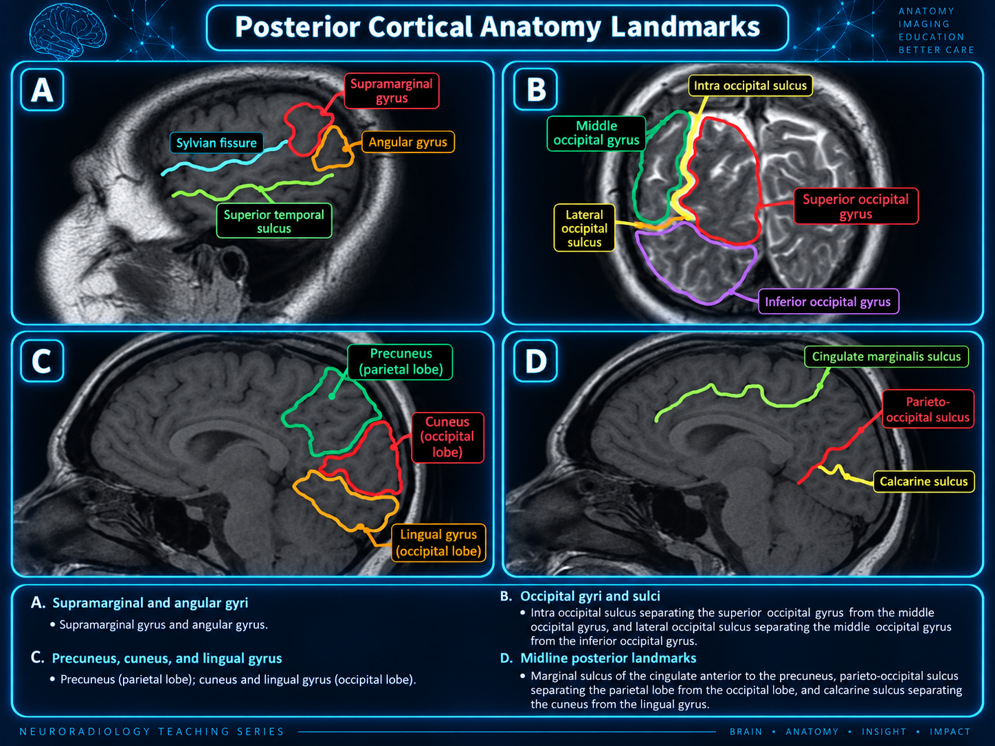
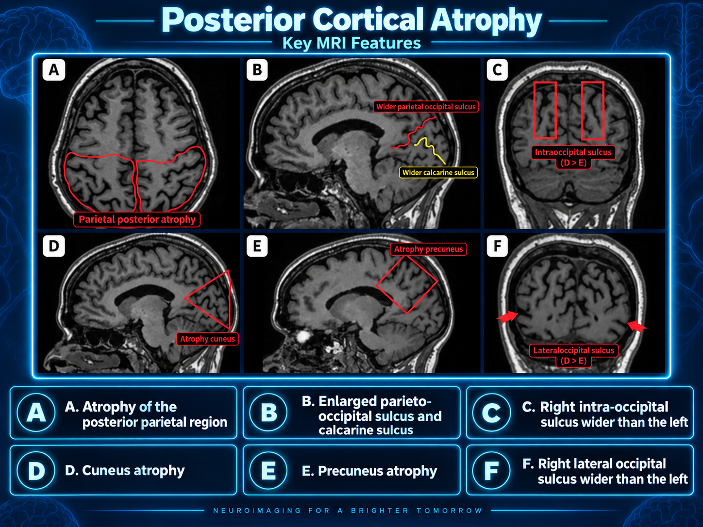
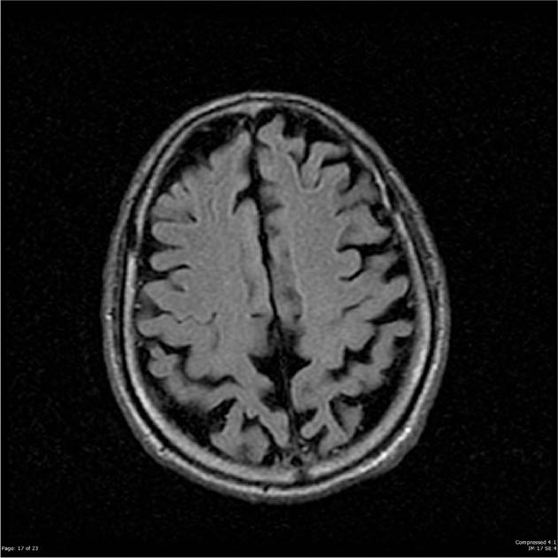
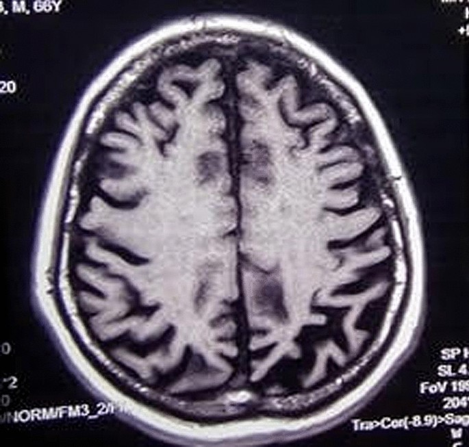
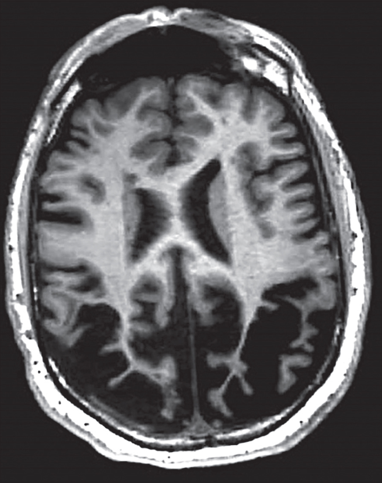
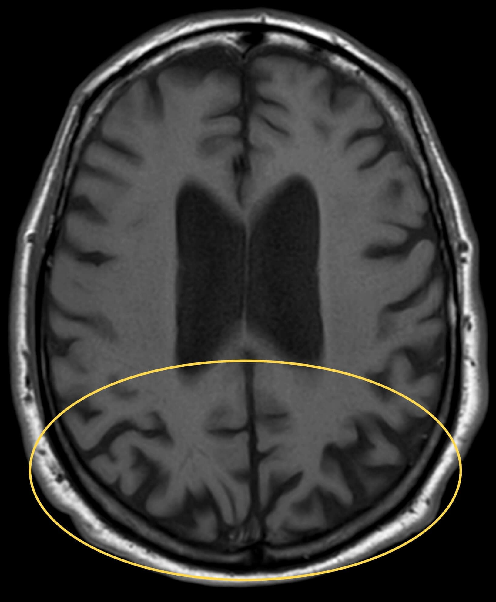
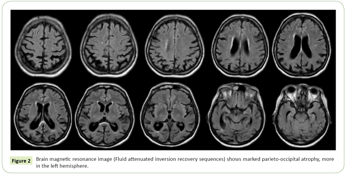
- Bilateral but often asymmetric (right > left) parietal and parieto-occipital atrophy
- Temporo-occipital involvement with relative preservation of medial occipital cortex
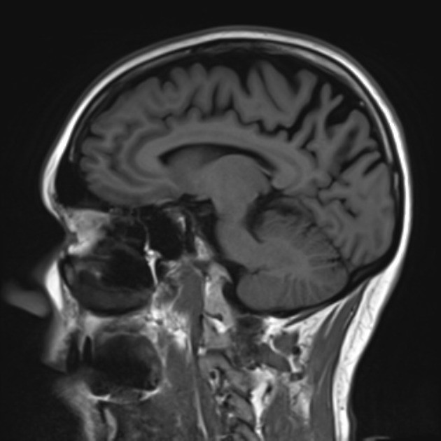
- Relatively preserved hippocampi distinguishing from typical Alzheimer disease
- Widened parieto-occipital sulcus compared to Alzheimer disease
Key Image Findings
- MRI shows bilateral but often right-predominant parietal and parieto-occipital atrophy as the core finding, with additional temporo-occipital involvement reflecting the clinical presentation of visual dysfunction.
- The parieto-occipital sulcus is wider in posterior cortical atrophy compared to typical Alzheimer disease, serving as a key distinguishing morphologic feature.
- Hippocampi remain relatively normal on MRI, which helps differentiate posterior cortical atrophy from typical Alzheimer disease where hippocampal atrophy is a prominent early finding.
- SPECT and PET imaging demonstrate hypoperfusion and hypometabolism in the parietal, parieto-occipital, and temporo-occipital regions that correlate with areas of morphologic atrophy.
- The 'occipital tunnel sign' on sagittal PET images shows asymmetric hypometabolism with relative preservation of medial occipital metabolism, representing a characteristic finding though not exclusive to posterior cortical atrophy.
- Functional imaging changes on SPECT and PET may precede morphologic atrophy on MRI, making nuclear medicine studies valuable for early detection.
- The pattern of atrophy is distinct from dementia with Lewy bodies, which shows frontal lobe and basal ganglia/thalami/midbrain involvement rather than predominant posterior involvement.
- CT imaging may allow gross volume changes to be appreciated but is less sensitive than MRI for detecting early atrophy patterns in this condition.
Differential Diagnosis
- Alzheimer disease: presents with older age at onset, more pronounced early memory loss, and absent or mild visuospatial features; shows prominent hippocampal atrophy and narrower parieto-occipital sulcus compared to posterior cortical atrophy.
- Dementia with Lewy bodies: characterized by frontal lobe and basal ganglia/thalami/midbrain atrophy rather than posterior predominance, with Parkinsonian symptoms later in disease course.
- Corticobasal degeneration: presents with limb rigidity, akinesia, dystonia, myoclonus, orobuccal/limb apraxia, cortical sensory deficits, and alien limb phenomenon rather than isolated visual dysfunction.
- Creutzfeldt-Jakob disease (CJD): can present with similar initial symptoms but has a much more rapid tempo of progression (typically months to 1-2 years versus 8-12 years) and distinctive diffusion restriction on DWI.
- Other pathologies in 'PCA-plus': dementia with Lewy bodies and corticobasal degeneration can coexist with posterior cortical atrophy presentation, requiring careful clinicopathologic correlation.
Discussion
Posterior cortical atrophy is recognized as a clinical syndrome with variable etiology rather than a single disease entity; while most cases represent a visual variant of Alzheimer disease with characteristic Alzheimer pathology (neuritic plaques and neurofibrillary tangles), other underlying etiologies including dementia with Lewy bodies, corticobasal degeneration, and prion disease can present identically.
The disease typically affects patients in their 50s-60s (earlier than typical Alzheimer disease) with no sex predilection, and the condition is likely underrecognized due to lack of general awareness and inconsistent diagnostic criteria.
Clinical presentation is dominated by disruption of higher-order visual processes resulting in visual agnosia, apraxia, prosopagnosia, alexia, and environmental disorientation; patients may develop Balint syndrome with simultanagnosia, oculomotor apraxia, and optic ataxia.
Early in disease course insight and episodic memory are relatively preserved, but generalized cognitive impairment including memory deficits develops as the disease progresses over 8-12 years.
Approximately 25% of patients develop visual hallucinations believed to result from complex interplay between midbrain, thalamus, and primary visual cortex rather than visual association areas, potentially representing a distinct clinical subgroup.
Gene mutations identified in posterior cortical atrophy include PSEN1, PSEN2, PRNP, GRN, and MAPT, suggesting shared genetic pathways with other neurodegenerative diseases.
Reporting Pearls
Describe the distribution precisely: "Bilateral parietal and parieto-occipital atrophy with asymmetric involvement (right > left), temporo-occipital changes, and relative preservation of hippocampi and medial occipital cortex." Compare the width of the parieto-occipital sulcus to differentiate from typical Alzheimer disease. Recommend correlation with PET/SPECT if available to document characteristic hypometabolism and 'occipital tunnel sign' supporting the diagnosis.
Pitfalls
- Do not mistake posterior cortical atrophy for typical Alzheimer disease based solely on clinical presentation; the younger age of onset (50s-60s), prominent visual symptoms, and preserved hippocampi are key distinguishing features on MRI.
- Avoid underrecognizing this condition due to its relative rarity; the pattern of right-predominant parieto-occipital atrophy with temporo-occipital involvement should raise suspicion even in patients with atypical presentations.
- Do not rely on MRI alone for diagnosis; remember that functional imaging (SPECT/PET) changes may precede morphologic atrophy and can demonstrate the characteristic 'occipital tunnel sign' and asymmetric hypometabolism essential for diagnosis confirmation.
- Be cautious with the diagnosis of isolated posterior cortical atrophy without considering PCA-plus variants; concurrent dementia with Lewy bodies or corticobasal degeneration features may be present requiring different management and prognosis counseling.