Progressive Supranuclear Palsy (PSP)
Key Images
Look For First
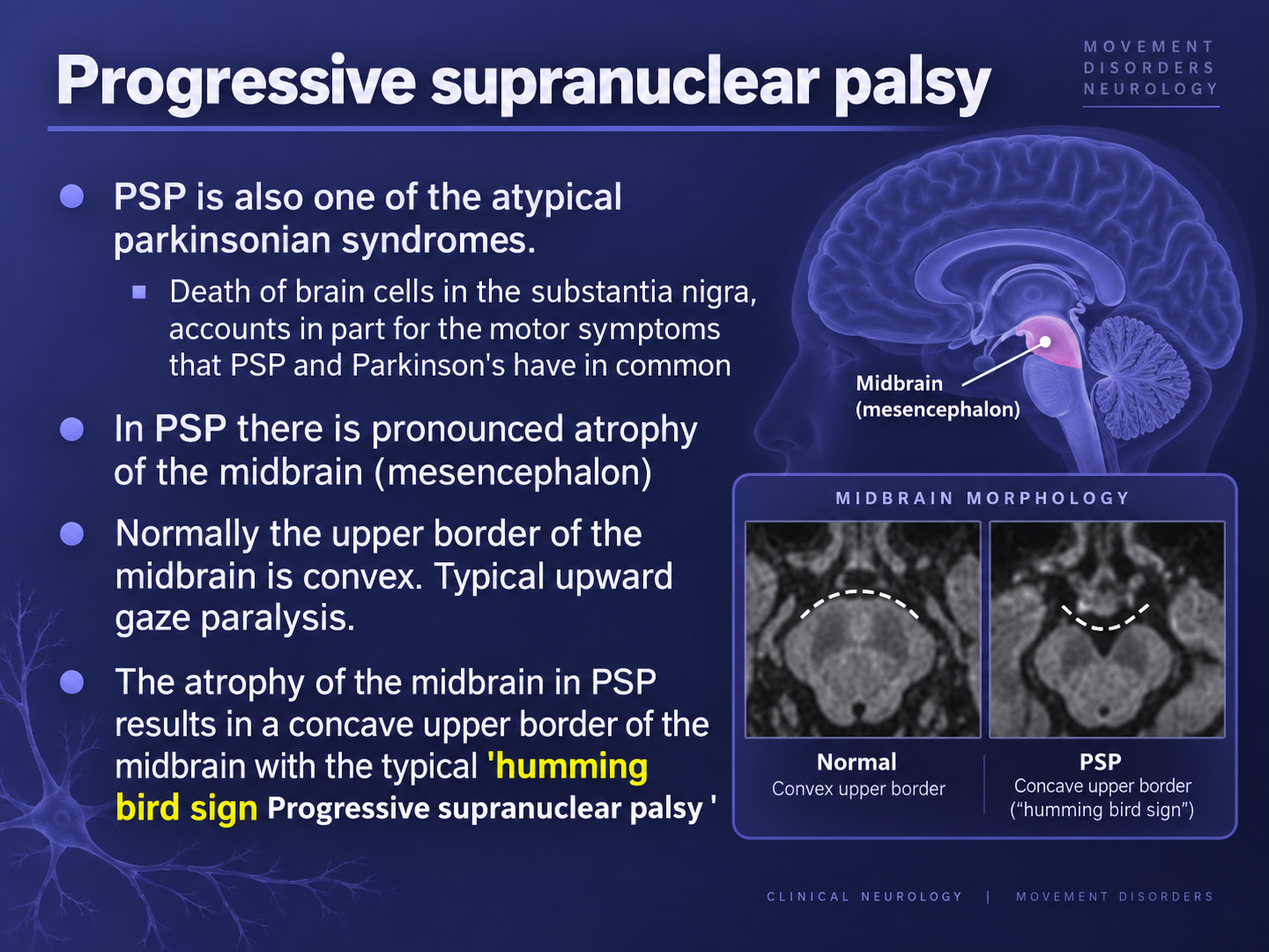
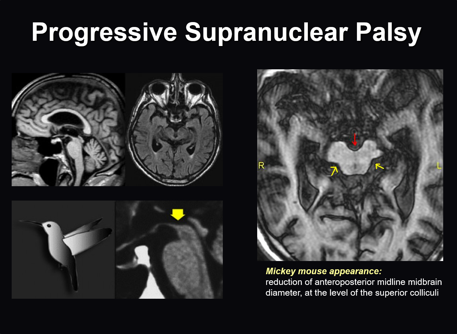
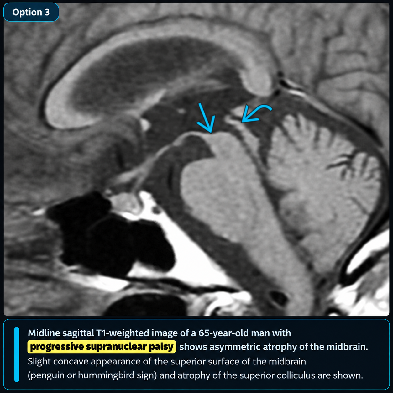
- Hummingbird or penguin sign on sagittal MRI: flattened or concave dorsal midbrain with preserved pons (100% specific)
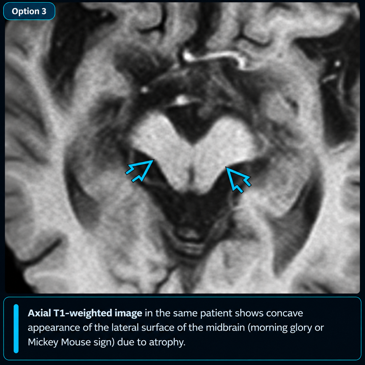
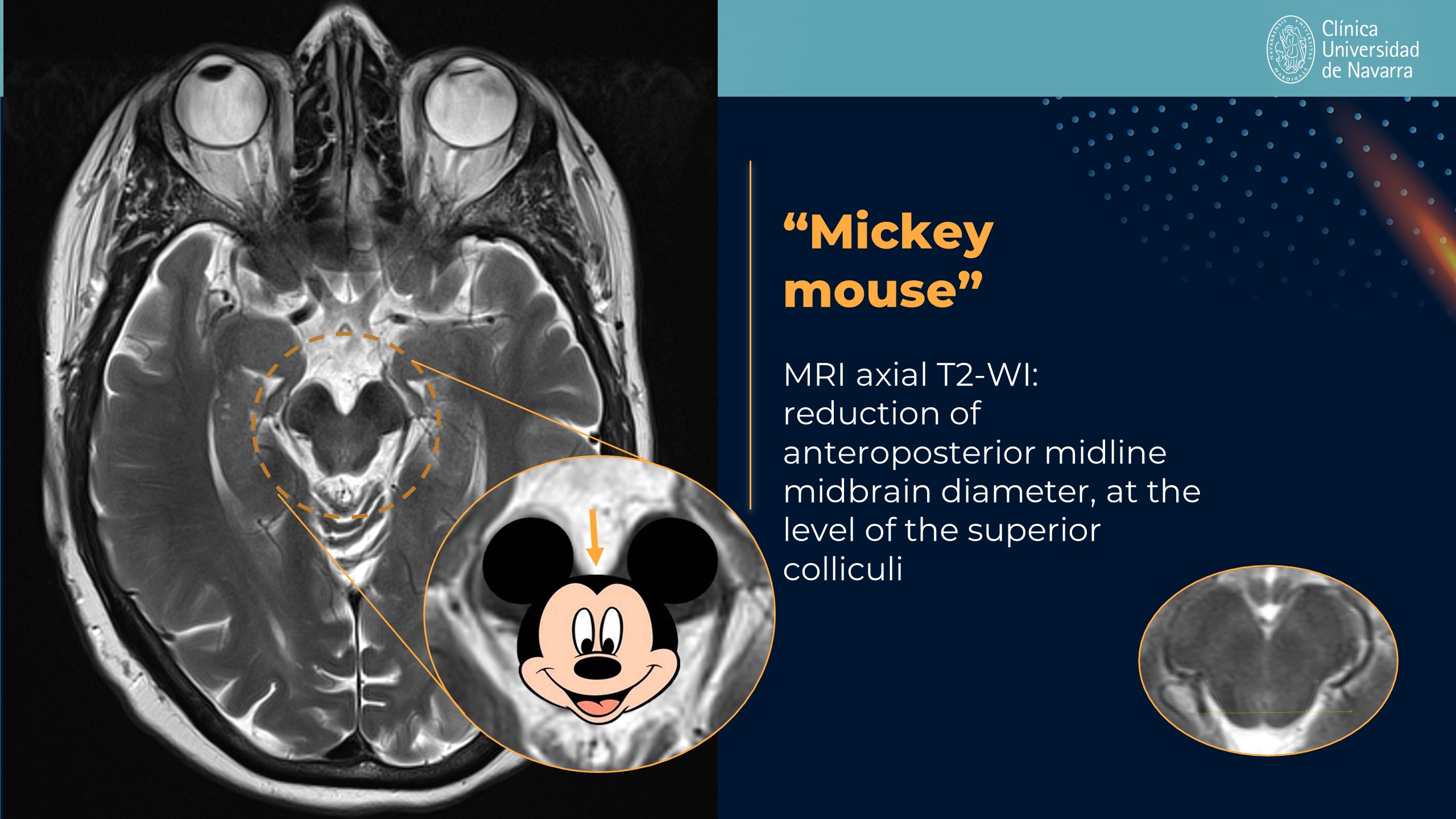
- Mickey Mouse sign on axial MRI: rounded peduncles with reduced anteroposterior midbrain diameter (<12 mm at superior colliculi)
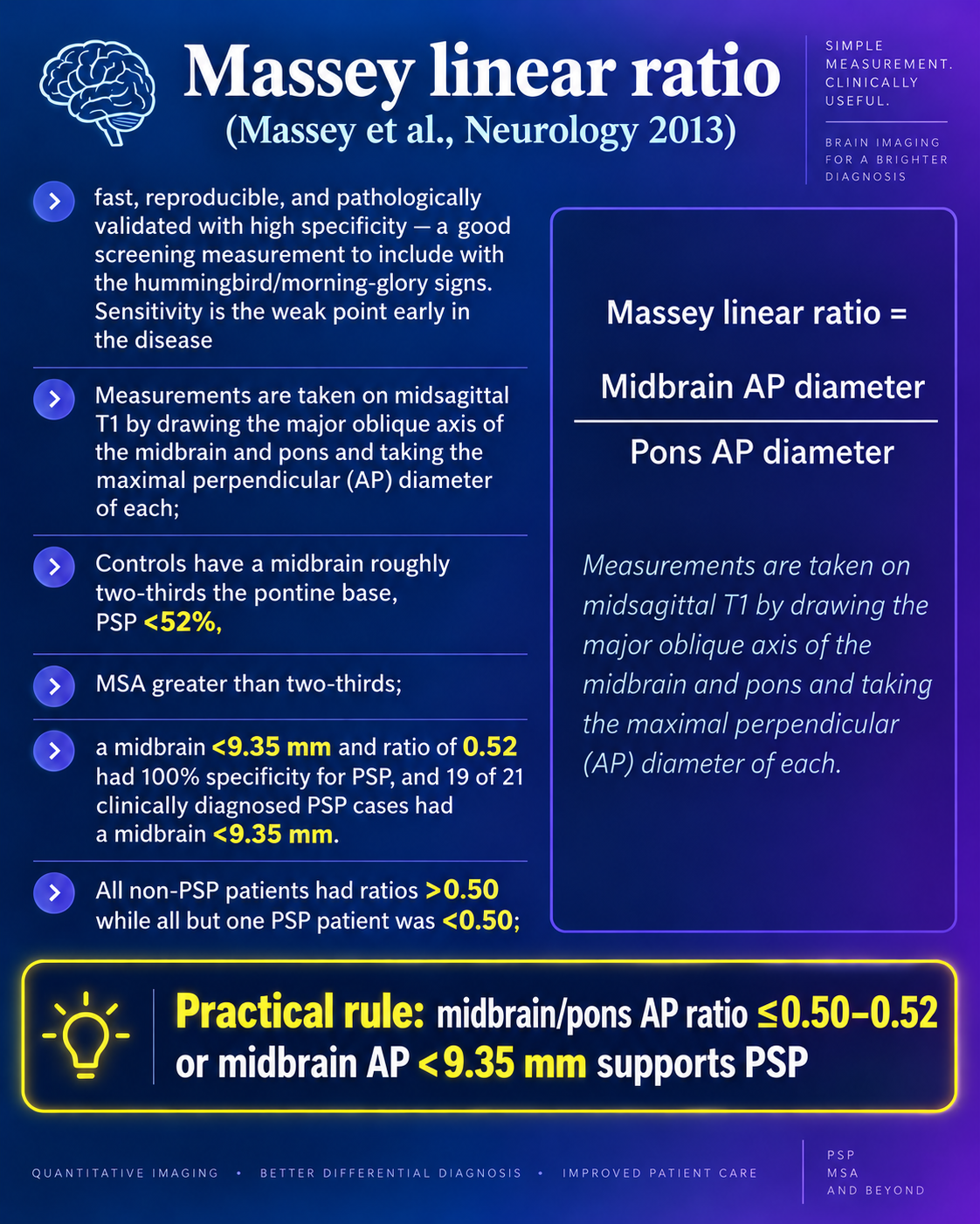
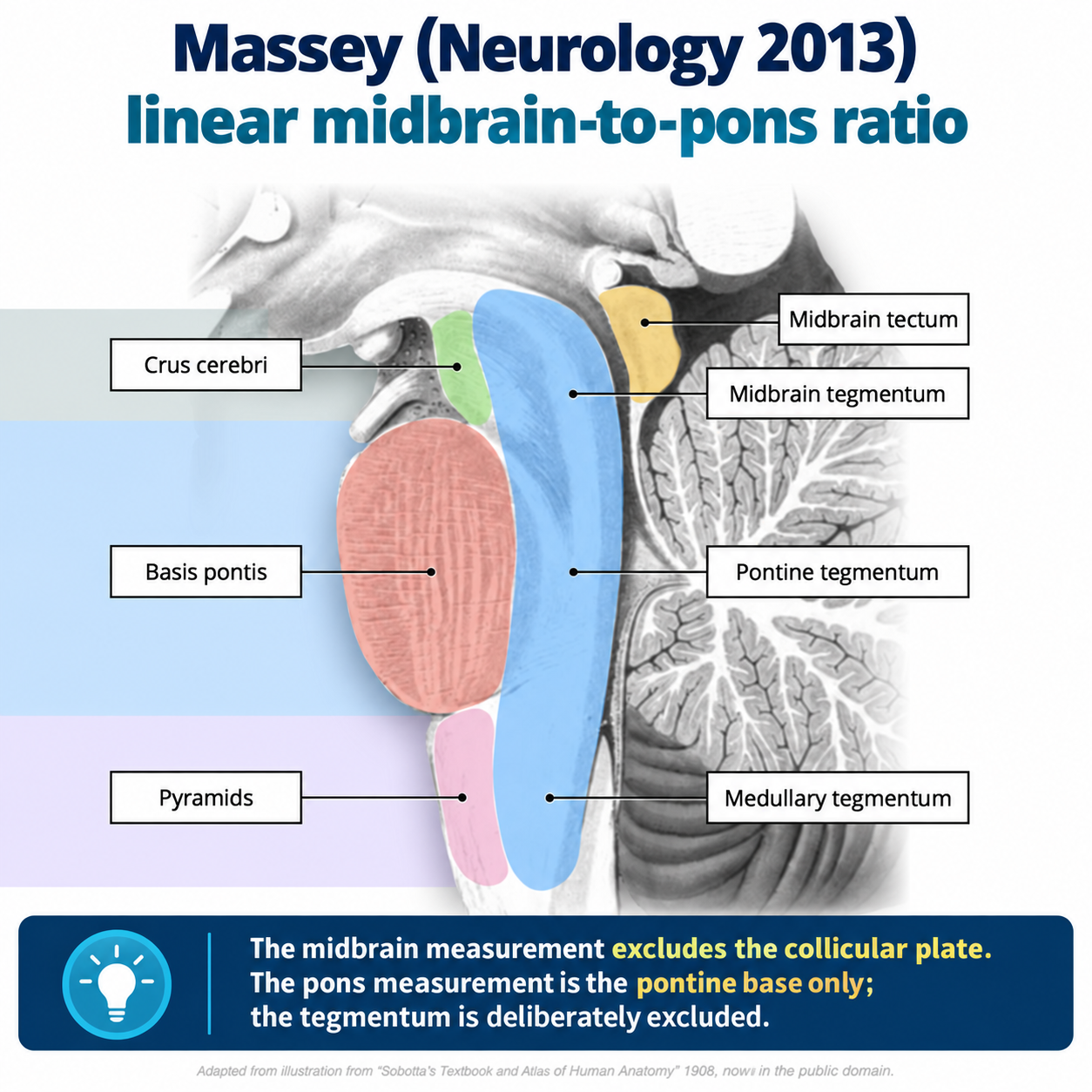
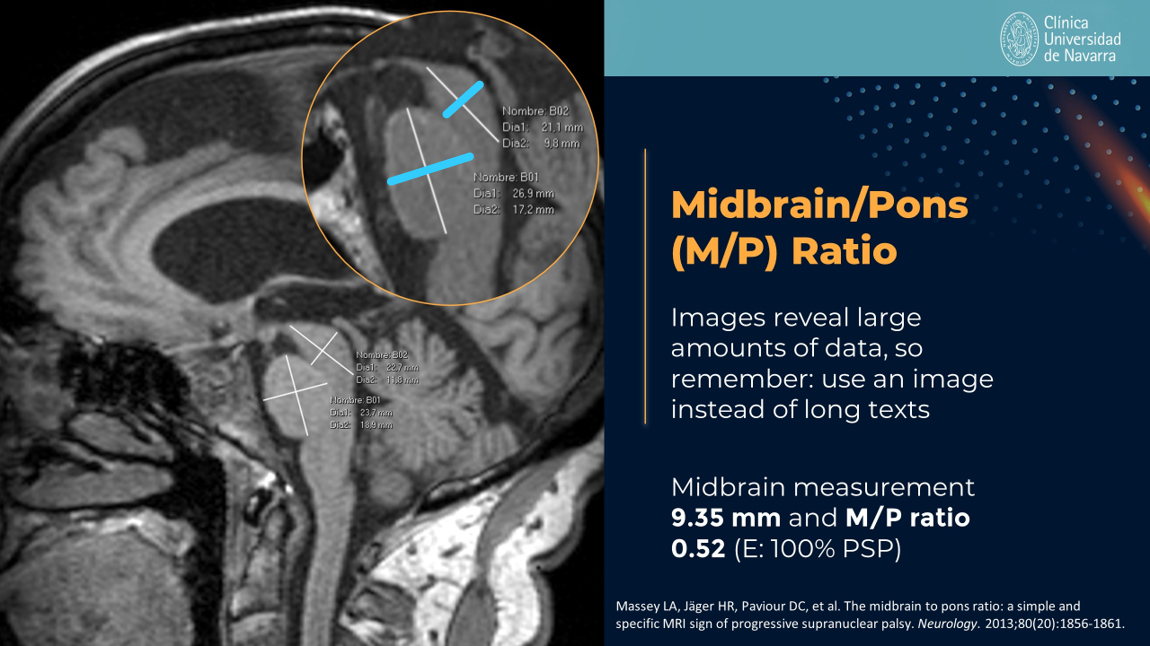
- Midbrain/pons area ratio <0.52: quantitative measure with 90% sensitivity and specificity that outperforms visual assessment
Key Image Findings
- Hummingbird/penguin sign: sagittal view shows characteristic flattening or concavity of the dorsal midbrain roof with dorsolateral tegmental hyperintensity on T2/FLAIR, while the pons remains relatively preserved; this sign is approximately 100% specific and 68-75% sensitive for PSP-RS.
- Mickey Mouse sign: axial image at the level of superior colliculi demonstrates rounded rather than oval peduncles with reduced anteroposterior midbrain diameter (<12 mm), achieving approximately 75% sensitivity for PSP.
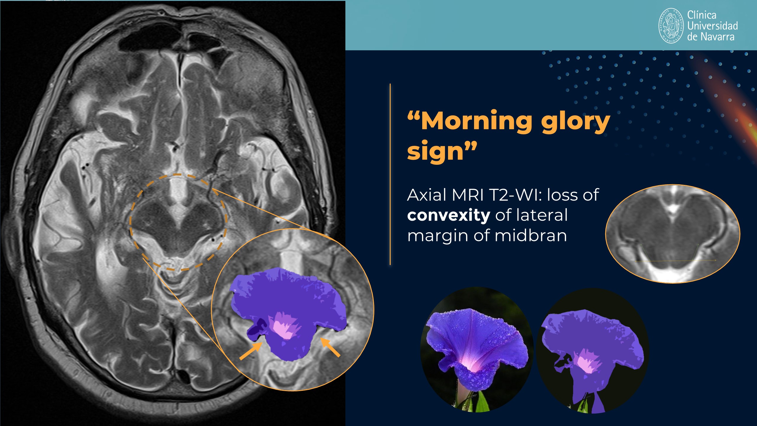
- Morning glory sign: axial loss of the normal lateral convexity of the midbrain tegmentum is 100% specific but only 45-50% sensitive, representing a helpful but less reliable finding.
- Midbrain/pons area ratio quantification: measurement on axial images at the midbrain level divided by pons area yields a ratio <0.52 with approximately 90% sensitivity and specificity, compared to normal values of ~0.24 and values >0.60 in Parkinson disease.
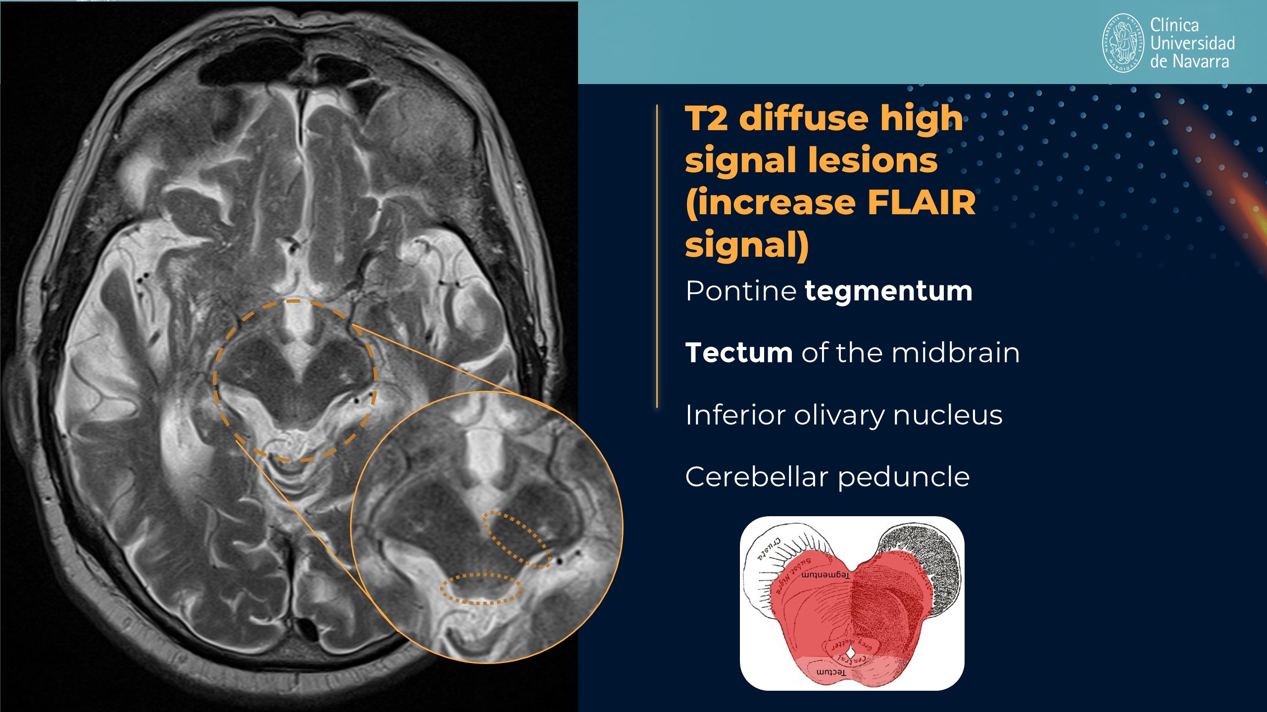
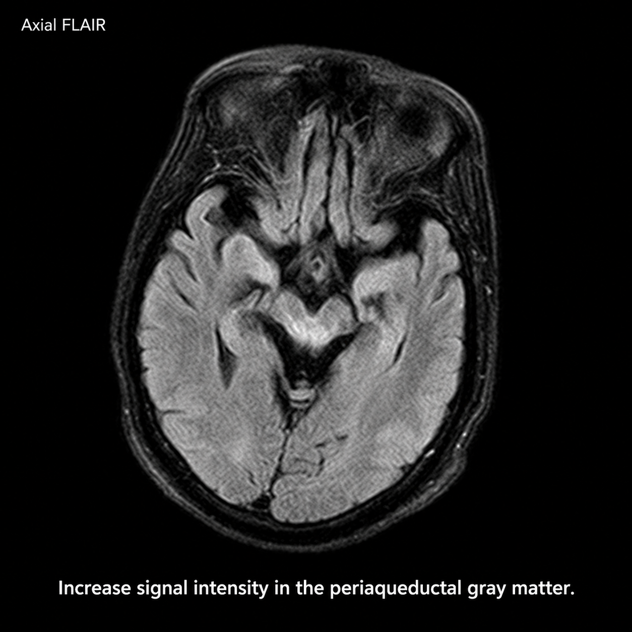
- T2/FLAIR hyperintensity in the pontine tegmentum, midbrain tectum, superior cerebellar peduncles (SCP), periaqueductal region, and inferior olive reflects the pathologic tau deposition and neuronal loss characteristic of PSP.
- Midbrain width measurement: absolute midbrain width <9.35 mm is highly sensitive (~85% in PSP-RS) and represents the single best planimetric measure for PSP detection on axial imaging.
- MRPI (MRI Parkinsonism Index) calculation as (pons/midbrain) × (middle cerebellar peduncle/SCP) yields a median value >19 in PSP versus 6.5 in MSA-P and 9.2 in controls, with near-100% accuracy in PSP-RS variants.
- Quantitative measures outperform visual signs in sensitivity and may precede the emergence of vertical gaze palsy by years, allowing early diagnosis before clinical manifestation of this hallmark feature.
Differential Diagnosis
- Parkinson disease: spares the midbrain and superior cerebellar peduncles, maintains normal midbrain/pons ratio >0.60, and shows preserved midbrain width on imaging.
- Multiple system atrophy-parkinsonism (MSA-P): shows atrophy of both pons and middle cerebellar peduncles with the characteristic hot-cross-bun sign; midbrain/pons ratio >0.52 and MRPI <10 helps distinguish from PSP.
- Corticobasal degeneration (CBD): features prominent asymmetric cortical atrophy and focal cortical ribboning rather than the selective brainstem atrophy pattern of PSP.
- Niemann-Pick type C: can mimic PSP with vertical supranuclear gaze palsy but typically shows additional findings such as cerebellar atrophy, white matter changes, and characteristic vertical supranuclear gaze palsy with downward gaze preference.
- Progressive supranuclear palsy variants: brainstem quantitative metrics are maximally abnormal in PSP-RS and PSP-frontal variants but near-normal in PSP-pure-gait-freezing, requiring recognition of phenotypic differences in imaging sensitivity.
Discussion
PSP is a 4R-tauopathy with pathologic hyperphosphorylated tau deposited in neurons and glia as globose tangles, tufted astrocytes, and coiled bodies, predominantly affecting the midbrain, basal ganglia, subthalamic nucleus, and frontal cortex, explaining the characteristic brainstem imaging findings.
Quantitative MRI measures (midbrain/pons ratio, MRPI, midbrain width) outperform visual qualitative signs in sensitivity and may detect pathologic changes years before the emergence of vertical supranuclear gaze palsy, enabling early diagnosis.
The hummingbird sign has exceptional specificity (~100%) for PSP-RS but moderate sensitivity (~70%), making it a highly specific but not sensitive sole diagnostic criterion; integration of quantitative measures improves detection.
Imaging findings show phenotype dependence with maximal brainstem atrophy in PSP-RS and PSP-frontal subtypes but near-normal brainstem metrics in PSP-pure-gait-freezing, requiring knowledge of variant presentations for accurate interpretation.
FDG-PET shows characteristic hypometabolism in the midbrain along with premotor, prefrontal, and anterior cingulate cortex involvement, with predominant midbrain hypometabolism qualifying as 'imaging-supported' PSP diagnosis.
DaT SPECT/PET demonstrates reduced striatal binding with loss of the normal comma shape, confirming neurodegenerative parkinsonism but not differentiating PSP from PD, MSA, or CBD; must be interpreted with structural MRI findings.
Reporting Pearls
When reporting PSP findings, describe the specific brainstem sign present (hummingbird, Mickey Mouse, or morning glory) with exact measurements: state the midbrain/pons ratio, absolute midbrain width, and MRPI value calculated as (pons/midbrain) × (MCP/SCP), noting that values <0.52, <9.35 mm, and >19 respectively support PSP diagnosis, then correlate with clinical presentation and note that quantitative metrics may precede gaze palsy emergence.
Pitfalls
- Relying on visual signs alone: the hummingbird and Mickey Mouse signs have only 68-75% sensitivity, so absence of these signs does not exclude PSP; quantitative midbrain/pons ratio and MRPI should always be calculated for improved diagnostic accuracy.
- Failing to measure quantitative metrics: midbrain/pons ratio, midbrain width, and MRPI outperform visual assessment and may detect PSP years before clinical gaze palsy emerges, so measurements should not be omitted even when qualitative signs appear normal.
- Over-reliance on brainstem metrics in PSP variants other than PSP-RS: in PSP-pure-gait-freezing and other phenotypes, brainstem metrics are near-normal on MRI so visual analysis has ≤50% sensitivity; these cases require higher clinical suspicion and may benefit from automated volumetric or machine-learning classifiers.
- Confusing PSP with MSA-P based on brainstem atrophy alone: both conditions show midbrain atrophy, but MSA-P also shows prominent pontine atrophy with the hot-cross-bun sign and preserved or elevated midbrain/pons ratio (>0.52), whereas PSP shows selective midbrain atrophy with ratio <0.52.