Perivascular spaces
Key Images
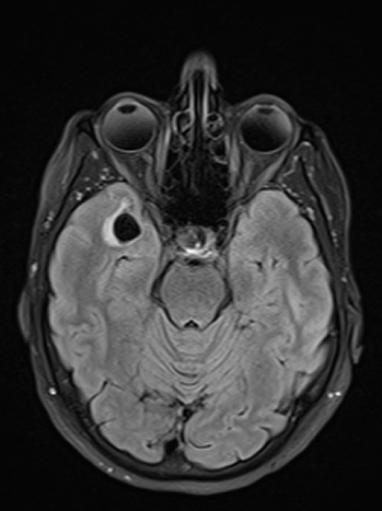
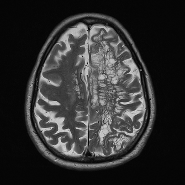
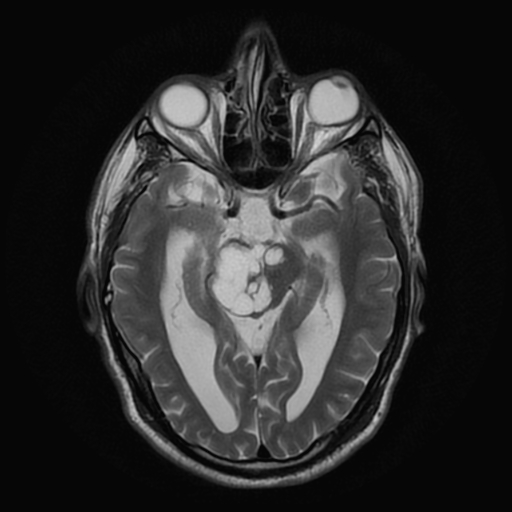
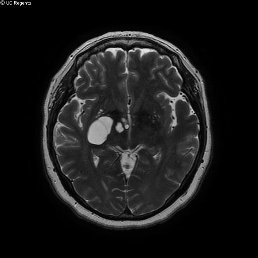
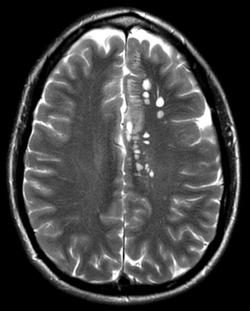
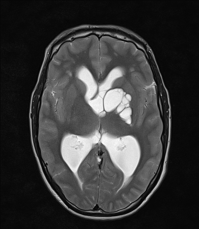
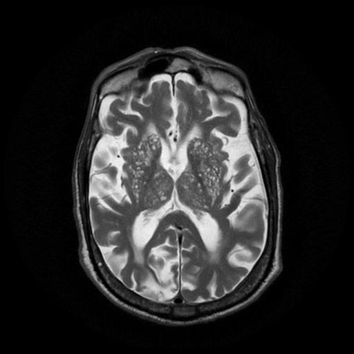
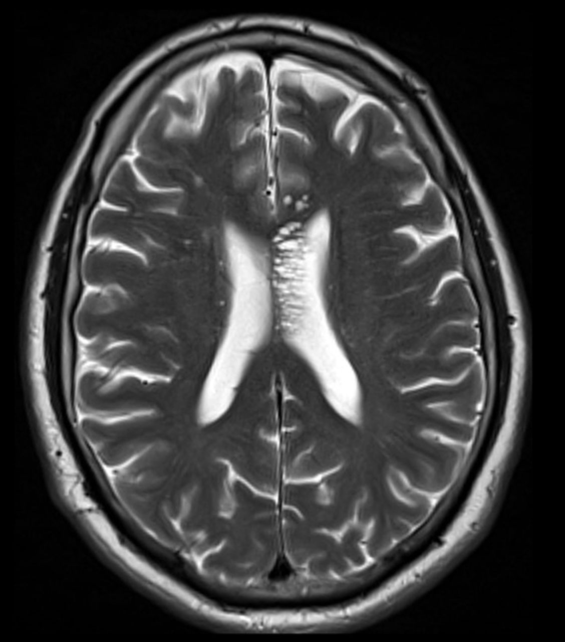
Look For First
- Linear or rounded CSF-signal fluid collections within basal ganglia along perforating arteries or in centrum semiovale, often <3 mm
- Traversing vessel sometimes visible through the space on T2 or SWI sequences
- Normal surrounding white matter without gliosis (unlike lacunar infarcts) to confirm benign nature
- Larger rounded/cystic morphology with possible thin T2-signal halo when >3 mm, helpful in distinguishing from other pathology
Key Image Findings
- Perivascular spaces are best seen on MRI and demonstrate CSF-identical signal on all pulse sequences (T1, T2, FLAIR, SWI)
- Most commonly visualized as thin linear regions <3 mm in maximum axial diameter when measured perpendicular to the traversing vessel, particularly in basal ganglia and centrum semiovale
- On T2 or SWI sequences, a traversing vessel is sometimes seen passing through the perivascular space, confirming the diagnosis
- As perivascular spaces enlarge, they develop a more rounded or oval cystic morphology, which may exceed 15 mm and become tumefactive with associated mass effect
- Surrounding white matter is typically normal, helping distinguish perivascular spaces from lacunar infarcts which show surrounding gliosis best seen on FLAIR
- CT demonstrates well-circumscribed fluid-density spaces with no enhancement or calcification; CT angiography may occasionally show a traversing vessel
- Anterior temporal lobe perivascular spaces are an exception to the normal white matter rule, as they may show increased T2/FLAIR signal related to associated vascular loops
- Large perivascular spaces may develop a thin increased T2-signal halo with positive mass effect, but maintain CSF signal centrally
Differential Diagnosis
- Lacunar infarcts—distinguished by surrounding gliosis on FLAIR, typically located in upper two-thirds of basal ganglia (perforating end-artery territory), and neutral or negative mass effect
- Choroid fissure cysts—consider in differential for small perivascular spaces
- Cystic neoplasms—distinguished by rare CSF-intensity on all MRI sequences and possible enhancement or mass effect
- Multiple sclerosis T1 black holes—can mimic small perivascular spaces but show different signal characteristics and clinical context
- Neurocysticercosis—identified by dot sign, enhancement, and calcification on imaging
- Non-neoplastic neuroepithelial cysts and ventricular diverticula—distinguished by location and morphology relative to ventricular system
Discussion
Perivascular spaces are normal anatomical structures consisting of invaginated pia and basement membrane surrounding small cerebral vessels; they function to transport CSF and metabolic waste from brain parenchyma
Enlargement of perivascular spaces visible on MRI is multifactorial in etiology, including parent arterial stiffening, protein aggregation (beta-amyloid), brain atrophy-related forces, and blood-brain barrier damage
Perivascular spaces are nearly ubiquitous on modern MRI due to improved resolution, seen in 50-100% of patients depending on defining criteria, with increasing prevalence in aging populations
Recent research suggests extensive basal ganglia perivascular spaces (état criblé) associate with cerebral small vessel disease changes and increased risk of subsequent dementia, though most remain asymptomatic
Perivascular spaces are classified into four types: type 1 (basal ganglia, lenticulostriate distribution), type 2 (cortical grey matter, medullary perforating arteries), type 3 (midbrain), and type 4 (anterior temporal lobe/insular)
Burden of perivascular spaces is highly heritable, especially in white matter, suggesting genetic predisposition independent of age-related factors
Reporting Pearls
Describe perivascular spaces by location (basal ganglia, centrum semiovale, etc.), morphology (linear vs. cystic), maximum diameter using STRIVE-2 criteria (<3 mm standard), and emphasize normal surrounding white matter and traversing vessels when visible to confirm benign etiology and distinguish from infarcts or cystic lesions requiring follow-up.
Pitfalls
- Mistaking lacunar infarcts for perivascular spaces by overlooking surrounding gliosis on FLAIR, which is present in infarcts but absent in perivascular spaces with normal white matter
- Confusing giant perivascular spaces (>15 mm) with cystic neoplasms; remember that true perivascular spaces maintain CSF signal on all sequences, whereas cystic neoplasms often show variable signal or enhancement
- Over-interpreting anterior temporal lobe perivascular spaces as pathologic when they show increased T2/FLAIR signal related to vascular loops; these represent a distinct entity
- Failing to identify a traversing vessel on high-resolution T2 or SWI sequences, which when visible is a key confirmatory finding for perivascular spaces rather than true cysts