Amyloid related imaging abnormalities (ARIA)
Key Images
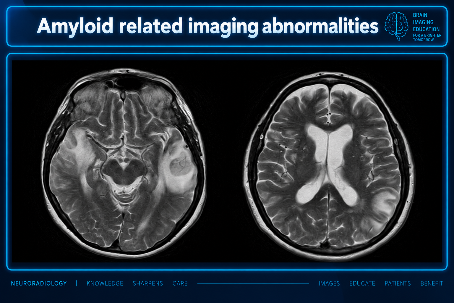
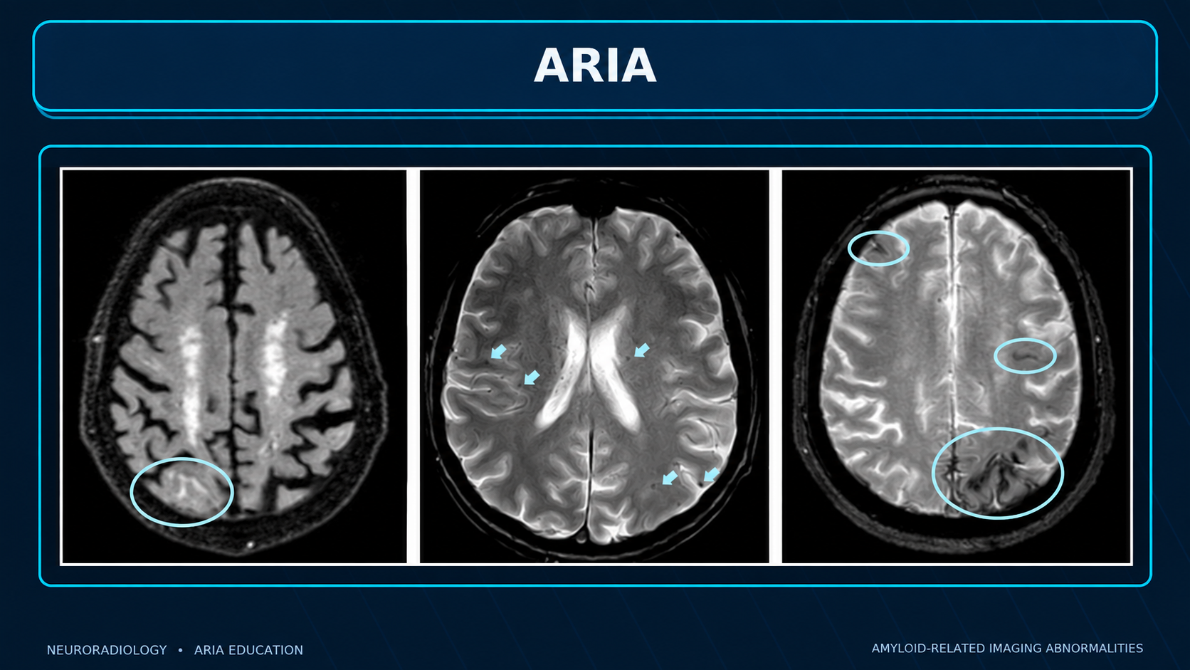
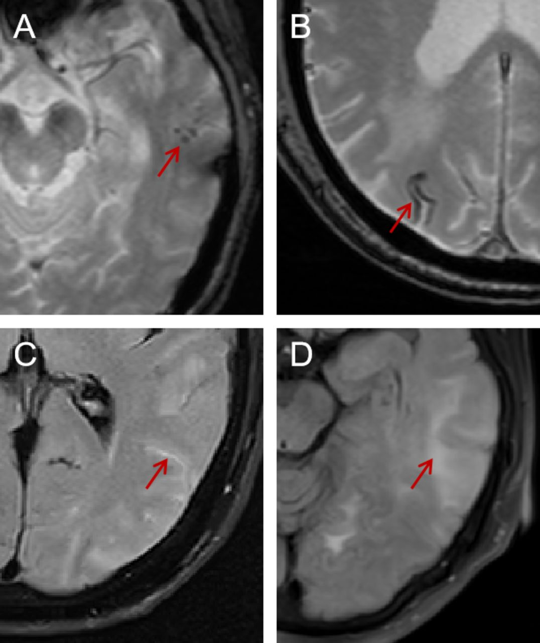
Look For First
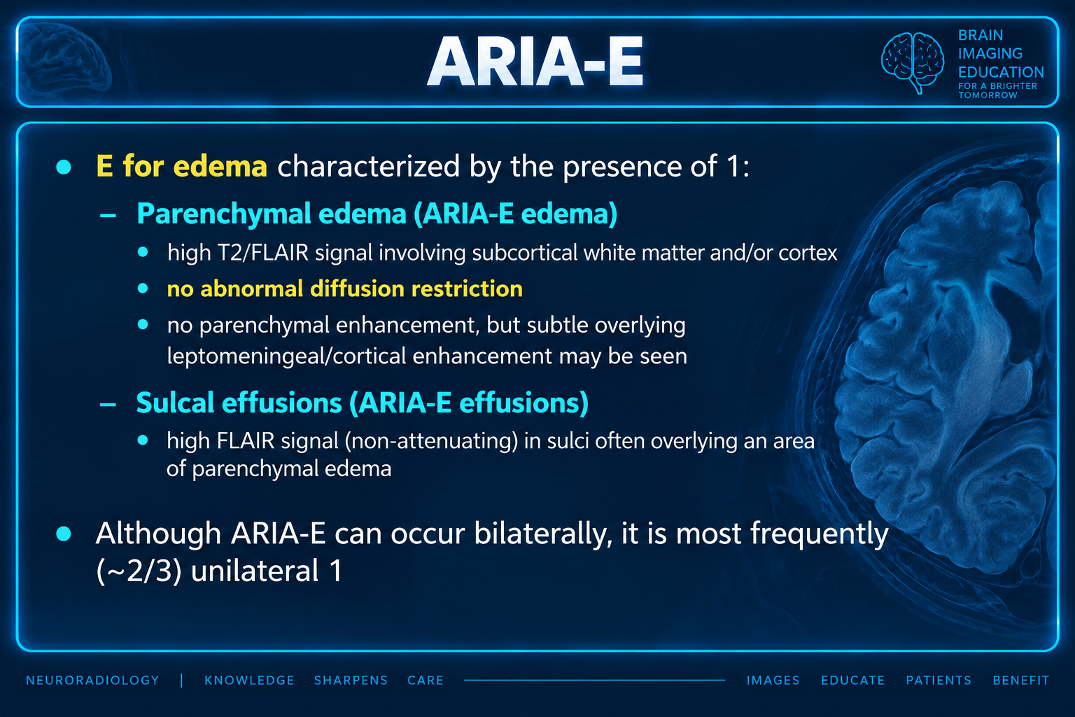
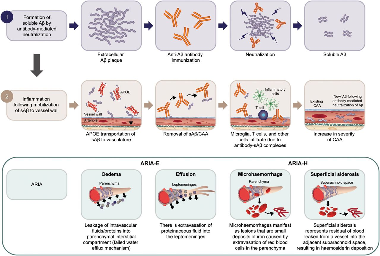
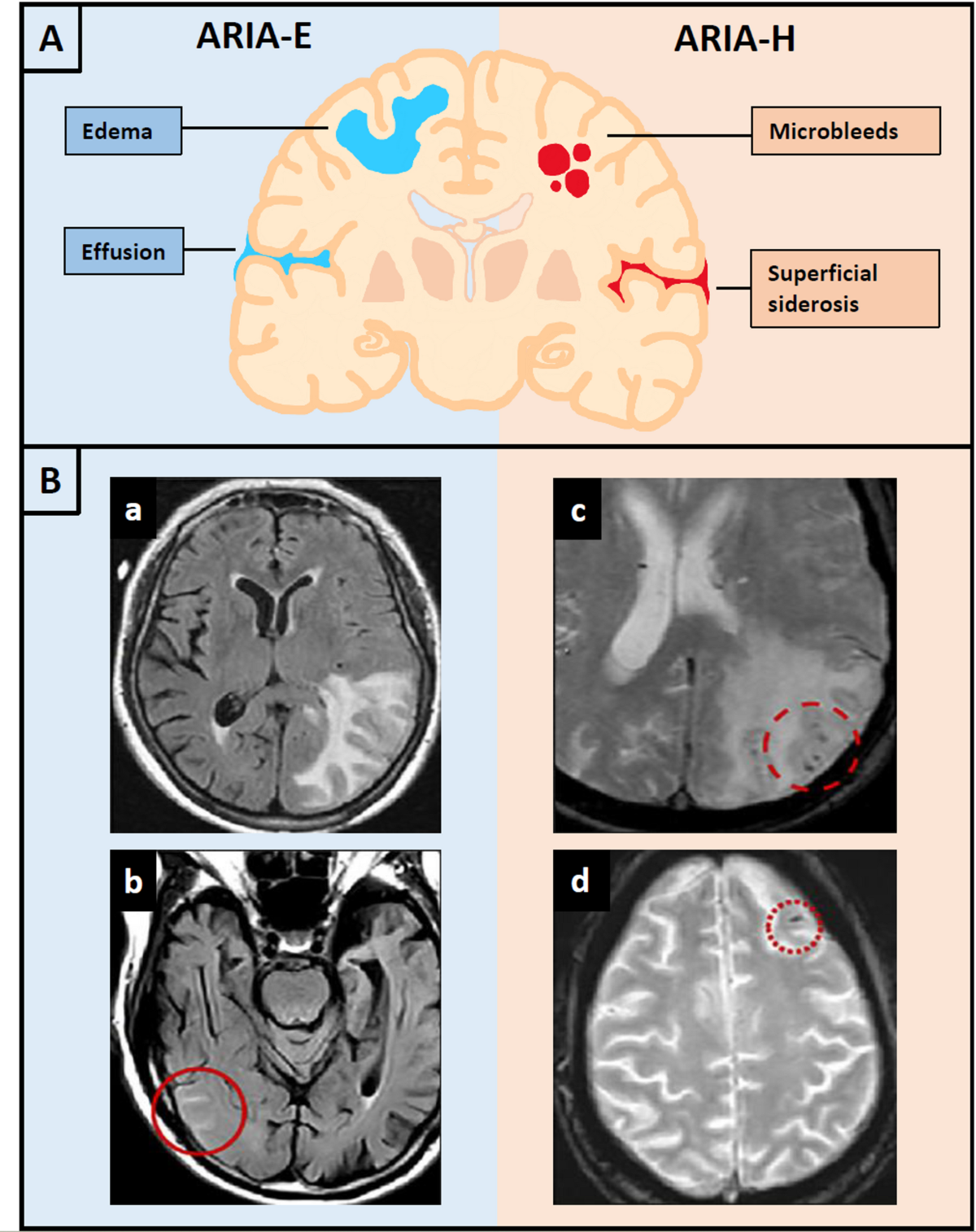
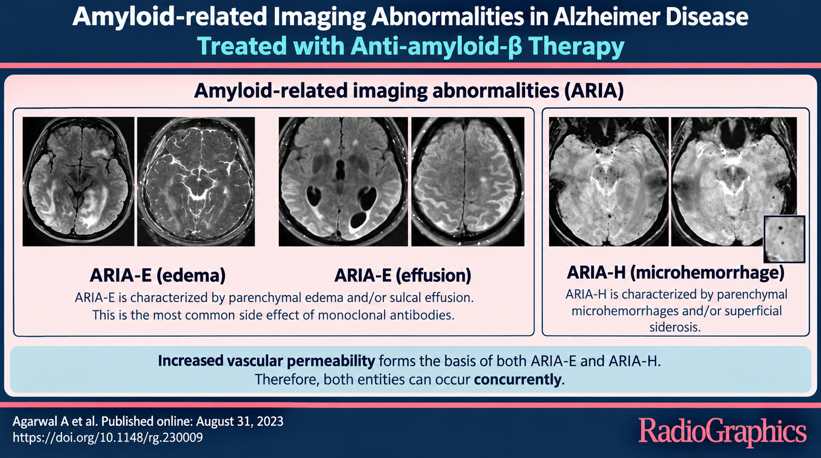
- High T2/FLAIR signal in subcortical white matter and/or cortex (ARIA-E parenchymal edema)
- High FLAIR signal in sulci overlying areas of parenchymal edema (ARIA-E sulcal effusions)
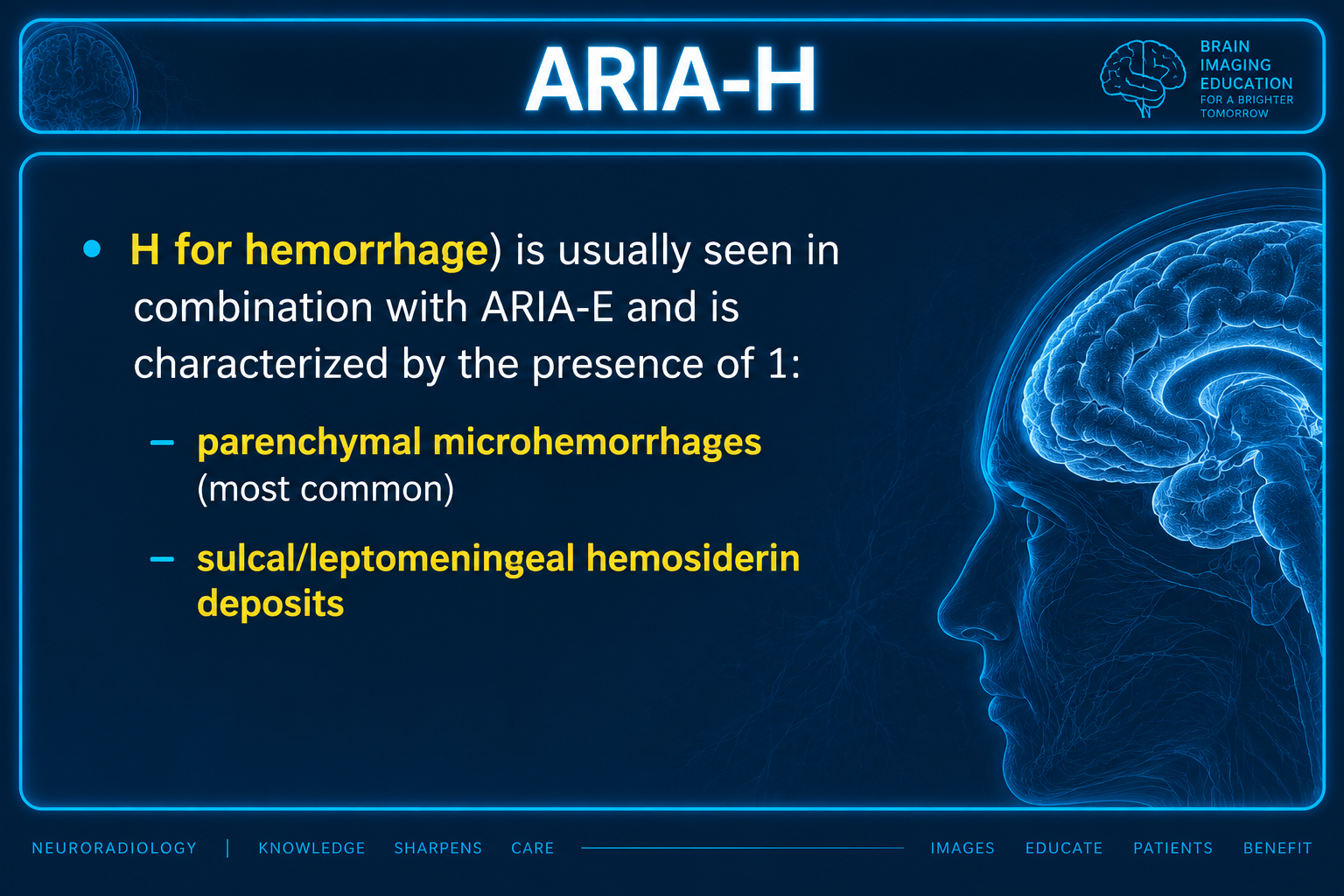
- Parenchymal microbleeds on T2* gradient echo sequences or cortical superficial siderosis (ARIA-H)
- Unilateral distribution of changes in approximately two-thirds of cases
Key Image Findings
- ARIA-E (edema) presents with high T2/FLAIR signal in subcortical white matter and/or cortex without abnormal diffusion restriction, distinguishing it from acute infarction.
- ARIA-E parenchymal edema may show subtle overlying leptomeningeal or cortical enhancement but lacks significant parenchymal enhancement, helping differentiate from inflammatory conditions.
- ARIA-E sulcal effusions appear as high FLAIR signal (non-attenuating) in sulci, often overlying an area of parenchymal edema, and can mimic subarachnoid fluid.
- ARIA-H (hemorrhage) is characterized by parenchymal microbleeds (most common) and sulcal/leptomeningeal hemosiderin deposits producing cortical superficial siderosis on susceptibility-weighted imaging.
- ARIA features frequently coexist as combined ARIA-E and ARIA-H, though they can occur independently; ARIA-H is usually seen in combination with ARIA-E.
- MRI is the primary modality for identifying ARIA, with FDA approval using T2* gradient echo sequences for monitoring, though more sensitive sequences like SWI are available.
- The distribution of ARIA changes correlates with the most active areas of amyloid removal, often appearing unilateral (two-thirds of cases) but can be bilateral.
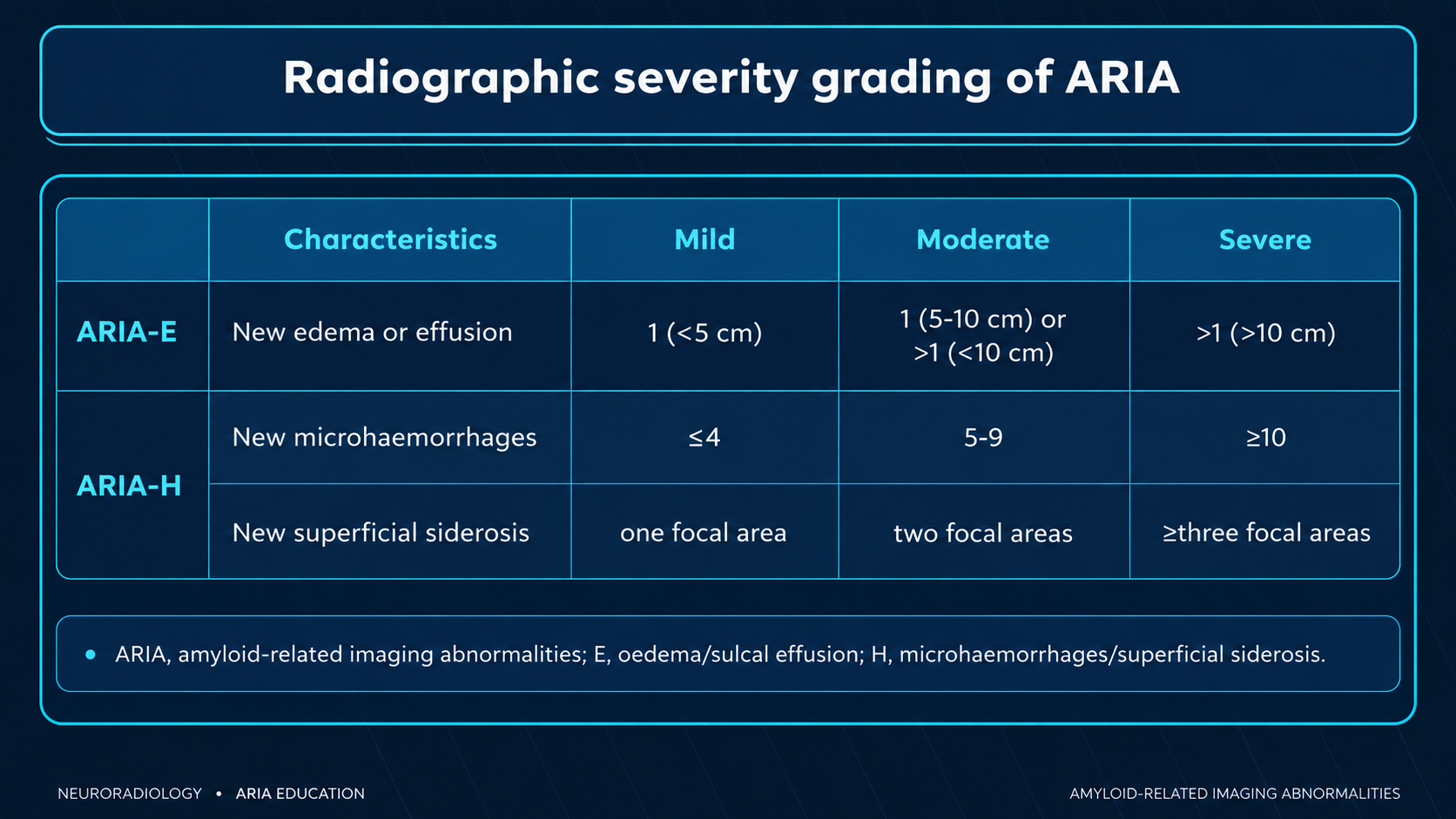
- ARIA-E severity is graded by FLAIR hyperintensity extent (mild <5 cm single location; moderate 5-10 cm or multifocal <10 cm; severe >10 cm), while ARIA-H is graded by microbleed count (mild ≤4; moderate 5-9; severe ≥10) or cortical superficial siderosis regions.
Differential Diagnosis
- Inflammatory cerebral amyloid angiopathy: imaging features are essentially identical to ARIA; clinical context is the distinguishing feature (occurs only in patients on anti-amyloid therapy for ARIA versus inflammatory pathology without anti-amyloid treatment).
- Posterior reversible encephalopathy syndrome (PRES): similar white matter edema usually in posterior distribution, can involve deep gray matter, typically associated with severe hypertension or other specific risk factors, and usually lacks microhemorrhages.
- Subarachnoid hemorrhage: sulcal effusions can mimic subarachnoid blood, distinguished by the high FLAIR signal (non-attenuating on CT) in ARIA versus attenuating blood on CT in true SAH.
- Meningitis: sulcal effusions can mimic sulcal pus accumulation, distinguished by clinical presentation (fever, meningeal signs), CSF abnormalities, and lack of parenchymal edema pattern seen in ARIA.
- Cerebral infarction: demonstrates diffusion restriction on DWI in acute phase and prominent enhancement in subacute phase, compared to absent DWI restriction and subtle enhancement in ARIA-E.
- Perilesional edema from mass lesions: edema surrounding metastases, primary tumors, or abscesses can appear similar; distinguished by the presence of an underlying enhancing lesion on contrast-enhanced imaging.
Discussion
ARIA pathophysiology involves breakdown of the blood-brain barrier from mobilization of amyloid previously deposited in blood vessels by anti-amyloid monoclonal antibodies.
Risk of ARIA varies significantly by drug type, with lecanemab showing lower rates (ARIA-E 13%, ARIA-H 17%), donanemab intermediate rates (ARIA-E 24%, ARIA-H 31%), and aducanumab showing highest rates (ARIA-E 35%, ARIA-H 20%) before its 2024 discontinuation.
Development of hemorrhagic lesions (ARIA-H) correlates with higher drug dose, presence of APOE ε4 alleles (same genetic risk factor for cerebral amyloid angiopathy), and concurrent use of antithrombotic agents requiring careful patient selection and monitoring.
ARIA is typically detected incidentally on imaging rather than from clinical symptoms, though some patients may present with headaches, vomiting, confusion, focal neurological deficits, or gait disturbance related to cerebral edema and irritation.
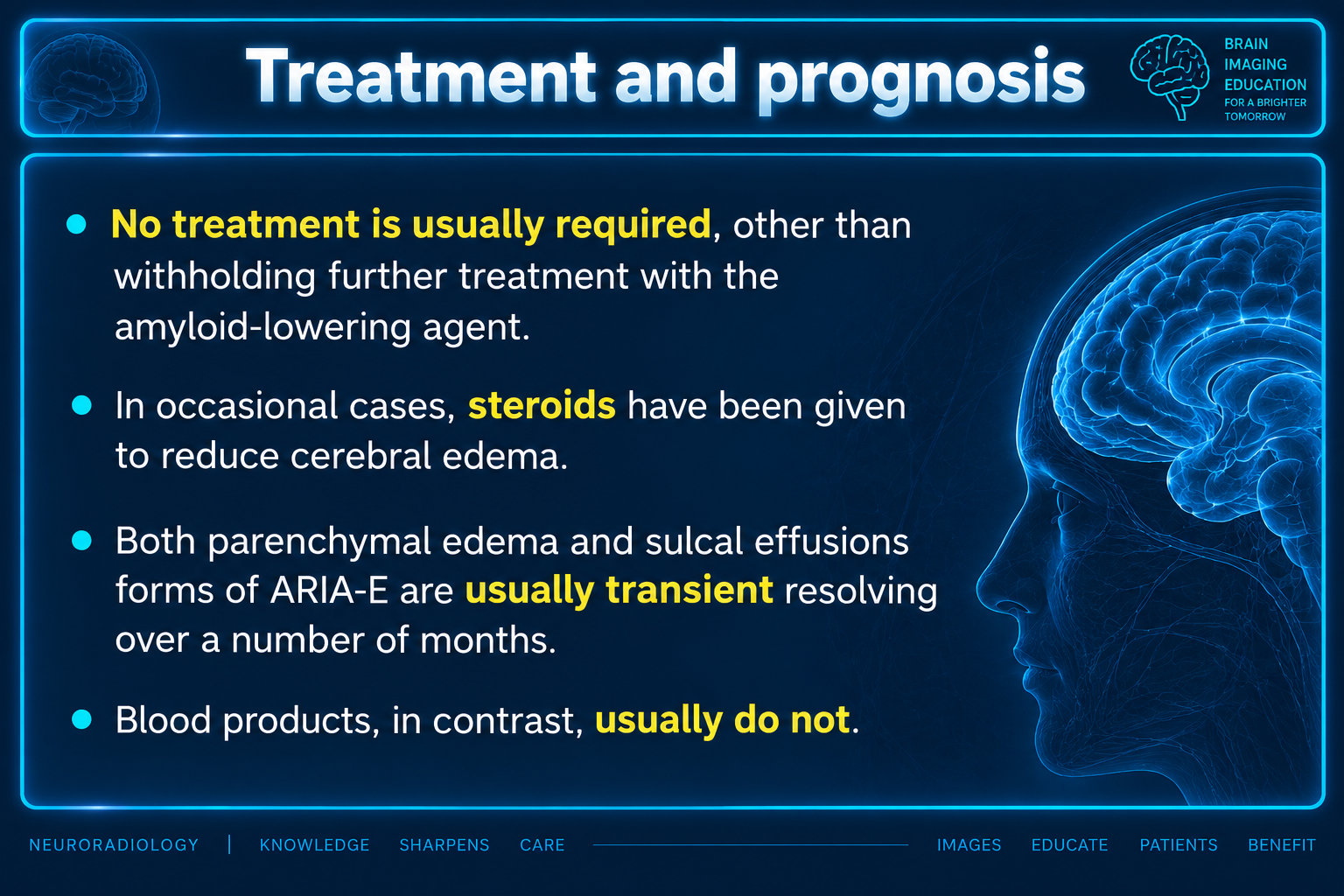
Most ARIA cases require no specific treatment beyond withholding further anti-amyloid therapy; however, severe cases with significant cerebral edema may benefit from corticosteroid administration to reduce swelling.
ARIA-E (edema and sulcal effusions) is usually transient, resolving over months, whereas blood products from ARIA-H typically do not resolve, requiring long-term monitoring and potentially permanent discontinuation of anti-amyloid therapy.
Reporting Pearls
Clearly specify the subtype (ARIA-E versus ARIA-H), quantify the extent of FLAIR hyperintensity with maximal dimension in centimeters, note the distribution (unilateral versus bilateral), describe the severity grade, and mention whether microhemorrhages or cortical superficial siderosis is present, as this standardized grading directly guides clinical decisions regarding continuation or discontinuation of anti-amyloid therapy.
Pitfalls
- Confusing ARIA-E sulcal effusions with acute subarachnoid hemorrhage: remember that sulcal effusions in ARIA are high FLAIR signal (non-attenuating on CT) and are usually associated with parenchymal edema, not diffuse subarachnoid blood.
- Misinterpreting ARIA-E parenchymal edema as acute infarction: ARIA-E lacks diffusion restriction on DWI/ADC sequences and shows absent or only subtle parenchymal enhancement, whereas acute infarcts show restricted diffusion and may show arterial enhancement.
- Under-recognizing ARIA-H microbleeds by using only conventional T2/T2* sequences instead of more sensitive susceptibility-weighted imaging (SWI), though FDA approval was based on T2* monitoring.
- Failing to correlate clinical context (current anti-amyloid therapy) with imaging findings, as inflammatory cerebral amyloid angiopathy can appear identical and requires different clinical management and prognostic counseling.