Ependymitis granularis
Key Images
Look For First
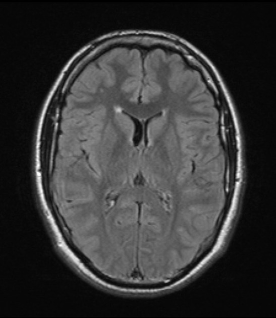
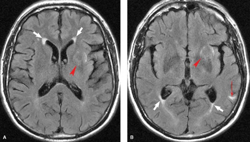
- Symmetrical foci of high T2/FLAIR signal anterior and lateral to the frontal horns
- Triangular morphology extending laterally from the callosal genu
- Small size, typically less than 1 cm, with normal low T1 signal intensity
Key Image Findings
- Ependymitis granularis appears as symmetrical periventricular foci of high T2 and FLAIR signal hyperintensity located anterior and lateral to the frontal horns.
- The lesions typically demonstrate a characteristic triangular morphology with apex directed laterally from the callosal genu.
- Size is usually less than 1 cm and lesions show normal or low signal intensity on T1-weighted imaging, reflecting no true pathology.
- The hyperintense signal on T2/FLAIR corresponds histologically to focal ependymal breakdown, astrocytic gliosis, decreased myelin, loose axonal organization, and increased periependymal fluid.
- The strictly symmetrical location anterior and lateral to the frontal horns is a key distinguishing feature that helps identify this variant.
- Unlike pathologic lesions, true ependymitis granularis has the small size and specific location that allows confident differentiation from genuine periventricular pathology.
Differential Diagnosis
- Transependymal edema: typically associated with enlarged ventricles and not confined to the frontal horns; represents true pathology requiring clinical correlation.
- Multiple sclerosis: periventricular lesions from MS are rarely symmetric, more ovoid in shape, and show predilection for perivenular distribution (ears of the lynx sign).
- Ears of the lynx sign: a very rare pattern seen in hereditary spastic paraplegia that can mimic ependymitis granularis but usually has additional neurologic findings.
- Chronic small vessel ischemia: produces periventricular changes that are usually asymmetric and often do not abut directly against the ventricular walls.
Discussion
Ependymitis granularis is purely an anatomical variant with no inflammatory etiology despite its name, contrary to a historical 1926 claim that linked it to chronic internal hydrocephalus.
Histologically, the lesions show focal ependymal breakdown, astrocytic gliosis, and decreased myelin with loose, random axonal organization that is smaller than adjacent normal white matter.
The increased periependymal and extracellular fluid contributes to the T2/FLAIR hyperintensity but does not represent true edema or pathology.
Recognition of the characteristic symmetrical, triangular, small morphology anterior and lateral to the frontal horns is essential to avoid misdiagnosis as demyelinating disease or ischemic change.
Clinical significance.
Usually incidental and asymptomatic: a normal correlate of aging and generalized cerebral atrophy in most cases.
Functional consequences when extensive: Because intact ependyma regulates fluid, ion, and small-molecule transport between parenchyma and CSF, widespread ependymal loss can impair this function and, with aqueductal involvement (e.g., mumps-related), cause acquired aqueductal stenosis and hydrocephalus.
Distinguish from true (infectious) ependymitis/ventriculitis, which is a clinically important, often severe process. Bacterial and fungal ependymitis are highly destructive, and infectious ventriculitis — associated with meningitis, ventricular catheters, or intraventricular abscess rupture — produces ependymal thickening, enhancement, and restricted-diffusion intraventricular debris on MRI, requiring CSF analysis and treatment. Granular ependymitis of aging is a separate, benign entity.
No clinical correlation or follow-up is needed when ependymitis granularis is confidently identified, as it is a benign incidental finding.
The distinction from pathologic lesions relies on size (>1 cm suggests pathology), asymmetry, and presence of T1 hypointensity in true lesions.
Reporting Pearls
Location and morphology (the most useful bedside discriminators)
Ependymitis granularis / periventricular caps:
- Smooth and symmetric.
- Well-marginated triangular “caps” at the anterior—and sometimes posterior—tips of the frontal or occipital horns.
- May appear as thin, “pencil-thin” linings or halos hugging the ventricular wall.
- Consistently begin in the ventricular horns and are essentially universal in the elderly.
- Correspond to Fazekas periventricular grade 1 (caps or pencil-thin lining).
Age-related (vascular) deep white matter disease:
- Punctate, early-confluent, or confluent foci in the deep and subcortical white matter.
- Often asymmetric and irregular.
- Not anatomically tethered to the ventricular horns.
- Always considered pathologic and associated with vascular risk factors.
Underlying pathophysiology (explains the imaging)
- Caps/periventricular hyperintensity (PVH): Reflects nonvascular change—ependymal lining disruption with subependymal gliosis, enlarged extracellular space, and transependymal CSF/interstitial fluid accumulation, likely from impaired glymphatic clearance rather than ischemia. Histology shows myelin pallor, ependymal thinning or discontinuity, and tortuous vessels without arteriolosclerosis. [1–3]
- Deep white matter hyperintensity (DWMH): Predominantly ischemic—arteriolosclerosis, microvessel stenosis, hypoperfusion, enlarged perivascular spaces, and, in more severe lesions, microinfarction with blood–brain-barrier breakdown and fibrinogen extravasation.
Pitfalls
- Mistaking ependymitis granularis for demyelinating disease (MS) if the symmetric, triangular morphology and location are not recognized; MS lesions are rarely symmetric and show different morphology.
- Over-interpreting the finding as representing true ependymitis or inflammation when it is purely an anatomical variant with no inflammatory process.
- Confusing larger or asymmetric lesions with ependymitis granularis; true pathologic lesions are >1 cm and show T1 hypointensity, whereas the variant is small and isointense on T1.
- Recommending unnecessary follow-up imaging for an incidental and benign finding, leading to patient anxiety and unnecessary healthcare cost.