Inflammatory cerebral amyloid angiopathy
Key Images
Look For First
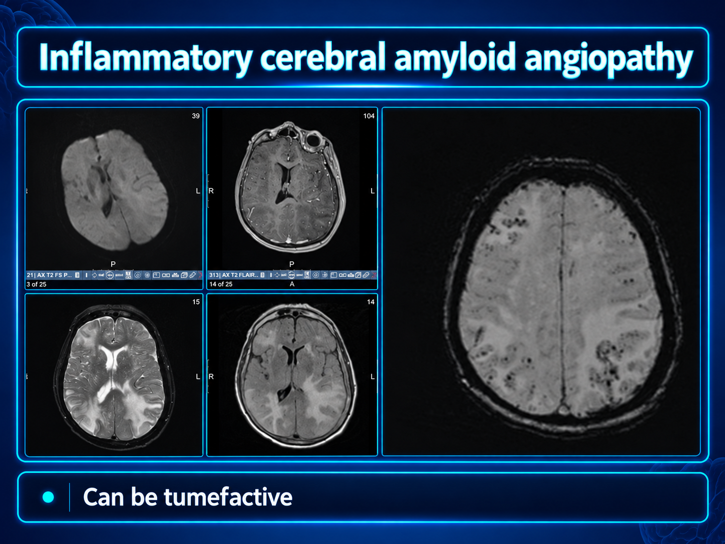
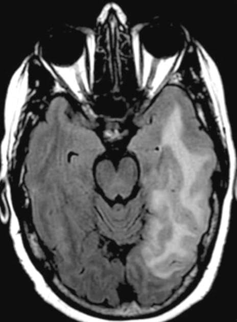
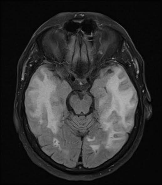
- Asymmetric subcortical white matter hyperintensities on T2/FLAIR that extend to immediately subcortical white matter with vasogenic edema
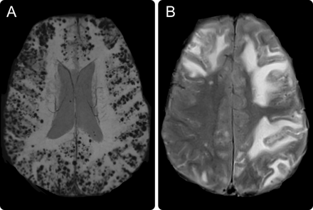
- Corticosubcortical hemorrhagic lesions on T2* gradient echo (microbleeds, macrobleed, or cortical superficial siderosis)
- Leptomeningeal enhancement on post-gadolinium T1 and/or sulcal hyperintensity on FLAIR adjacent to white matter changes
Key Image Findings
- MRI white matter lesions appear as T2/FLAIR hyperintensities in subcortical white matter (unifocal in most cases, multifocal in ~30%) representing vasogenic edema that may show localized mass effect with gyral swelling
- Hemorrhagic lesions are demonstrated as signal loss on T2*-weighted sequences (susceptibility weighted or gradient echo) and present in approximately 90% of cases, including cerebral microbleeds that are usually located in the same areas as T2/FLAIR high signal and are more numerous than in noninflammatory CAA
- Cerebral microbleeds in inflammatory CAA do not follow the prevalent occipital pattern typical of noninflammatory CAA but are distributed more broadly throughout the brain
- Cortical involvement of white matter lesions is encountered and predisposes to seizures, distinguishing this entity from pure deep white matter disease
- Leptomeningeal involvement detected through leptomeningeal contrast enhancement on T1 C+ (Gd) (seen in 50-70% of patients) and/or sulcal hyperintensity (non-nulling) on FLAIR representing subarachnoid involvement
- Leptomeningeal enhancement and sulcal hyperintensities are usually topographically adjacent to white matter changes and gyral swelling
- MRA and vessel wall imaging may show medium-sized artery involvement with multifocal stenoses and wall thickening/enhancement, though wall enhancement is not specific for inflammation
- CT imaging demonstrates subcortical white matter changes as areas of low density with localized mass effect, but MRI is the modality of choice due to superior visualization of microhemorrhages and cortical superficial siderosis
Differential Diagnosis
- Noninflammatory cerebral amyloid angiopathy: distinguished by lack of inflammatory changes, asymmetry, leptomeningeal involvement, and different microbleed distribution (occipital predominance in noninflammatory vs. multifocal in inflammatory)
- Amyloid-related imaging abnormalities (ARIA) from monoclonal antibody therapy: occurs in specific clinical context of amyloid-lowering treatment rather than spontaneous disease
- Primary angiitis of the central nervous system (PACNS): distinguished by involvement of large- and medium-sized vessels on angiography with vasculitis features, whereas iCAA spares large vessels on DSA
- Stroke from other causes: distinguished by lack of multifocal vasogenic edema, hemorrhagic lesions at different distribution, and absence of leptomeningeal involvement
- Viral meningoencephalitis: differentiated by clinical presentation, leptomeningeal enhancement pattern, and absence of characteristic corticosubcortical hemorrhages and white matter changes of CAA
- Demyelinating disease (ADEM, MS): lacks the hemorrhagic component and specific distribution of vasogenic edema in subcortical white matter extending to immediately subcortical regions
Discussion
Inflammatory cerebral amyloid angiopathy is distinguished from noninflammatory CAA by the presence of perivascular and intramural inflammatory infiltrates, fibrinoid deposits, and granulomatous changes affecting both leptomeninges and cortical vessels as confirmed by brain biopsy with Congo red staining and beta-amyloid immunoreactivity
The diagnostic criteria for 'probable' iCAA require asymmetric white matter hyperintensities on T2-FLAIR extending to immediately subcortical white matter (not from past hemorrhage), while symmetric lesions only meet criteria for 'possible' iCAA
At least one corticosubcortical hemorrhagic lesion (macrobleed, microbleed, or cortical superficial siderosis) is required for both probable and possible diagnoses, present in approximately 90% of cases
Clinical presentation is typically acute or subacute with symptoms including headache, decreased consciousness, behavioral change, focal neurological signs, seizures, or acute cognitive decline that cannot be attributed to acute intracerebral hemorrhage alone
The distinction between cerebral amyloid angiopathy-related inflammation (CAA-ri) with perivascular-only inflammation and amyloid β-related angiitis (ABRA) with vessel wall involvement is not reliably predicted on imaging and may require pathological correlation via biopsy
Leptomeningeal enhancement is seen in 50-70% of patients and may be the predominant radiographic finding in some pathologically-confirmed cases with minimal white matter or hemorrhagic lesions on imaging, particularly in amyloid β-related angiitis presentations
Reporting Pearls
Describe the combination of asymmetric subcortical white matter hyperintensities on T2/FLAIR extending to immediately subcortical regions, with multiple corticosubcortical microbleeds on T2* gradient echo, leptomeningeal enhancement on post-gadolinium T1, and sulcal hyperintensity on FLAIR as characteristic of inflammatory cerebral amyloid angiopathy, noting the topographic relationship between white matter changes, hemorrhagic lesions, and leptomeningeal involvement.
Pitfalls
- Confusing inflammatory cerebral amyloid angiopathy with noninflammatory CAA by missing the asymmetry and leptomeningeal involvement features that distinguish the inflammatory form
- Failing to recognize that microbleeds in inflammatory CAA are more numerous and multifocal compared to the occipital-predominant pattern of noninflammatory CAA, potentially leading to mischaracterization of the underlying pathology
- Misinterpreting vessel wall enhancement on MRA or vessel wall imaging as diagnostic of vasculitis when wall enhancement is not specific for inflammation and does not establish large/medium vessel vasculitis (which is absent in iCAA as shown by normal DSA)
- Overlooking prominent leptomeningeal involvement as a primary manifestation, particularly in amyloid β-related angiitis cases where leptomeningeal changes may predominate over typical white matter lesions, leading to consideration of meningitis or other leptomeningeal processes instead of iCAA