Larynx Anatomy

Imaging orientation: Review the larynx from the hyoid through the inferior cricoid, identifying the supraglottis, glottis, and subglottis in sequence. On every examination, assess the mucosal surfaces, cartilage framework, pre-epiglottic and paraglottic spaces, anterior and posterior commissures, and the expected symmetry of the vocal cords.

Visual Reference

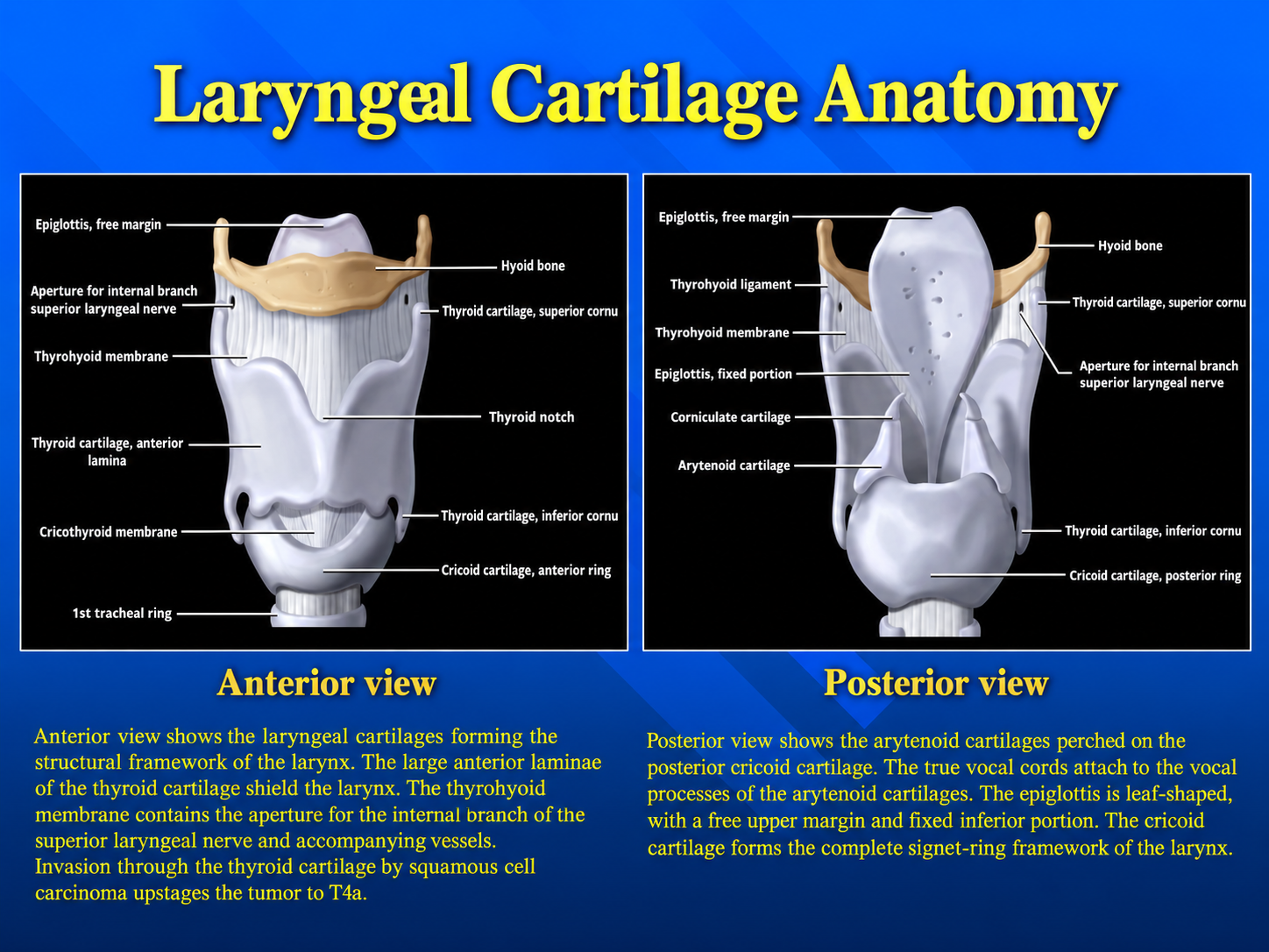
Anterior and posterior views of the laryngeal cartilages
Cartilaginous FrameworkClick to enlarge
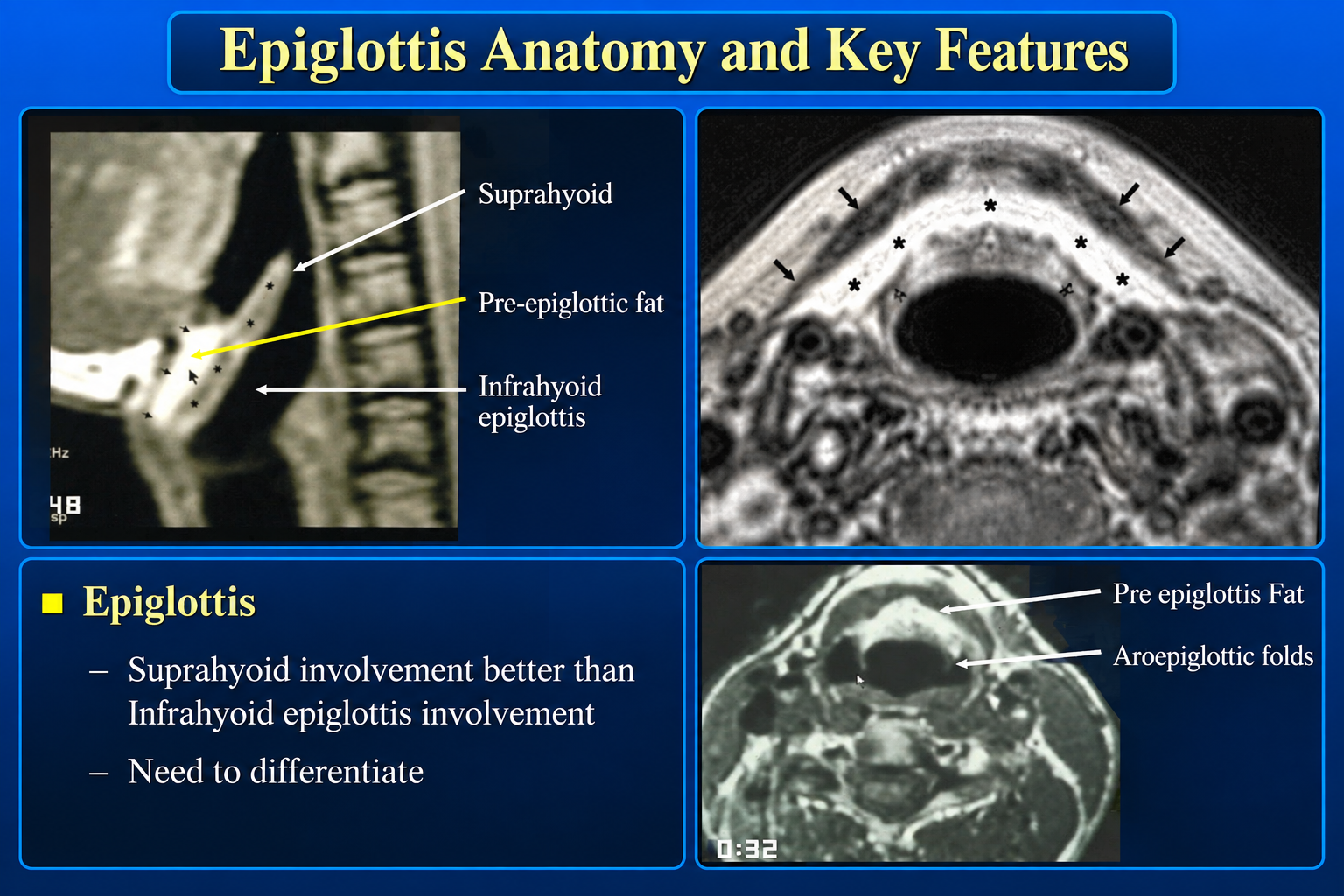
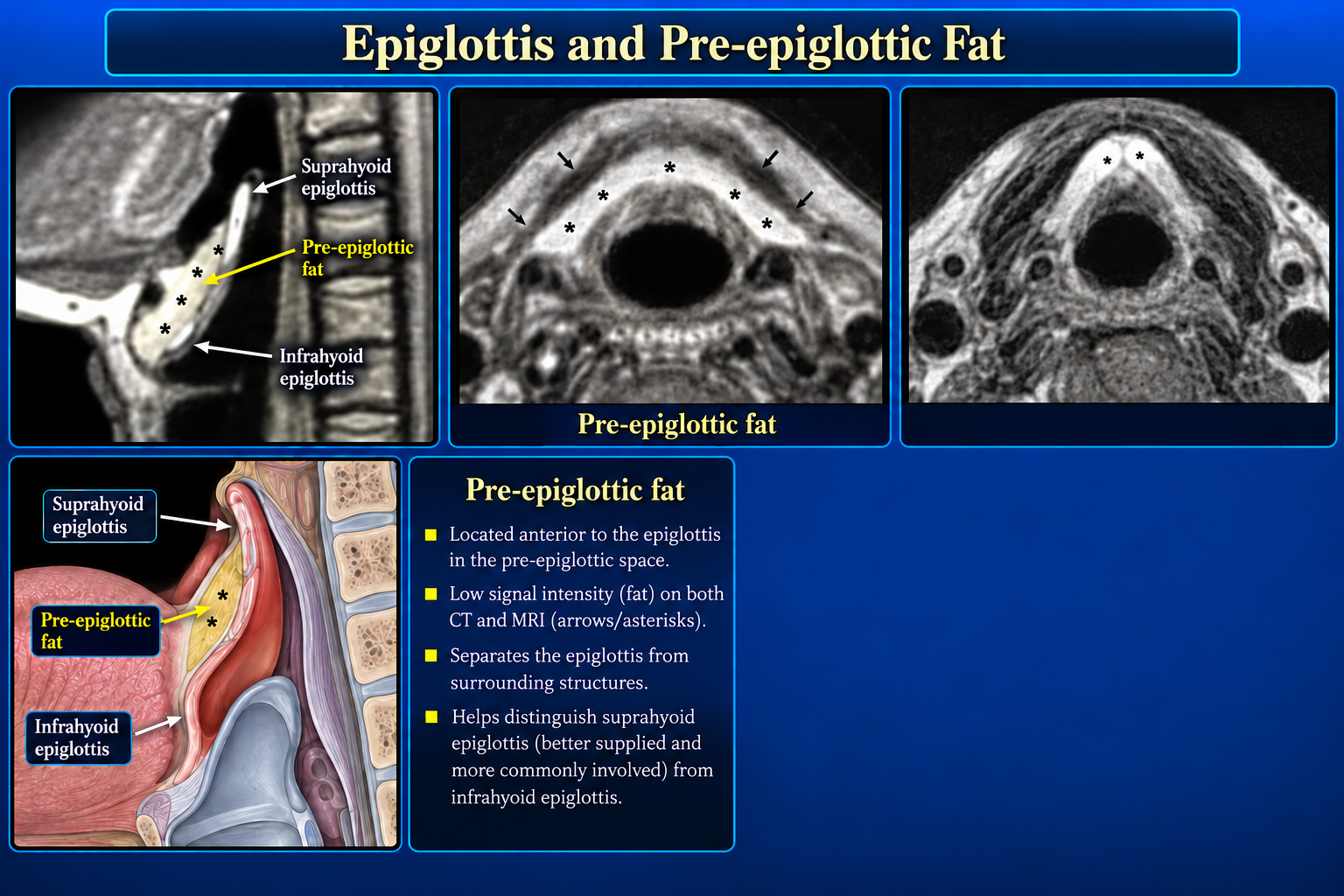
Epiglottis and pre-epiglottic fat on sagittal and axial imaging
Epiglottis and Pre-epiglottic FatClick to enlarge
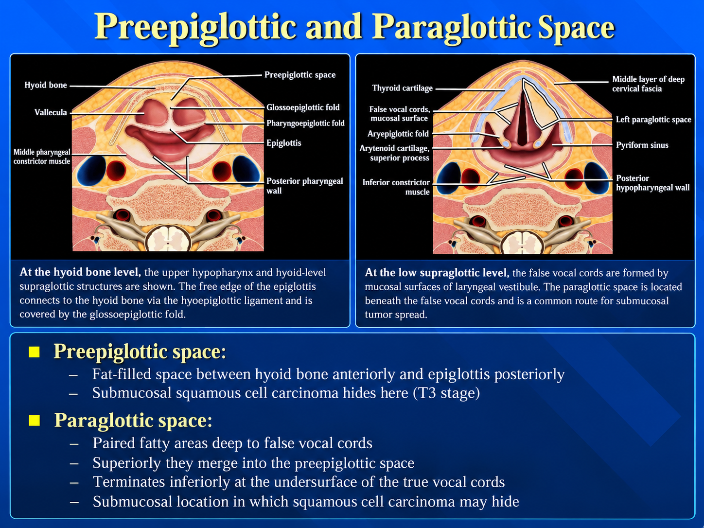
Preepiglottic and paraglottic spaces
Deep Laryngeal SpacesClick to enlarge
Epiglottis anatomy and key imaging features
Epiglottic LandmarksClick to enlarge
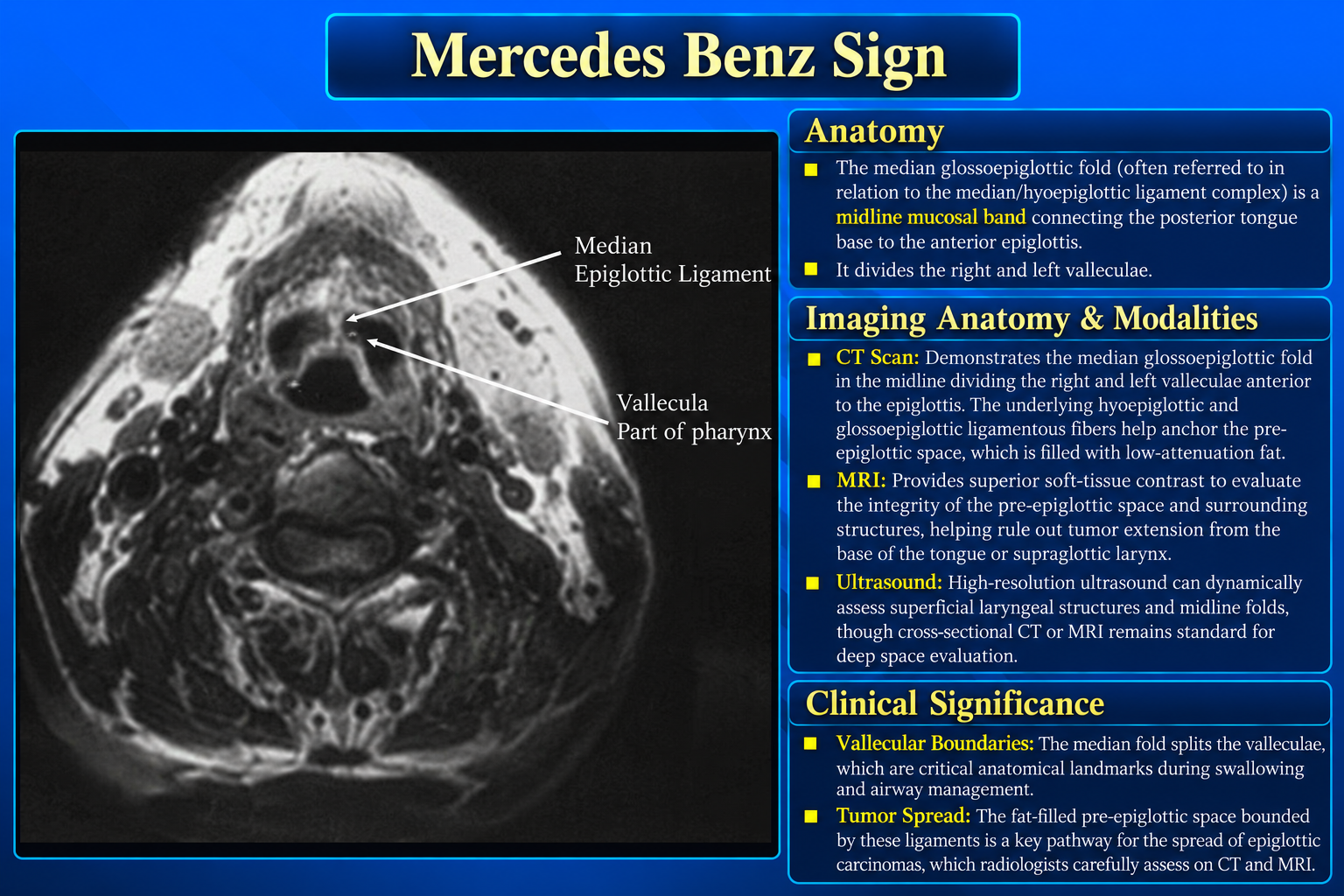
Median glossoepiglottic fold and vallecular anatomy
Vallecular Mercedes-Benz SignClick to enlarge
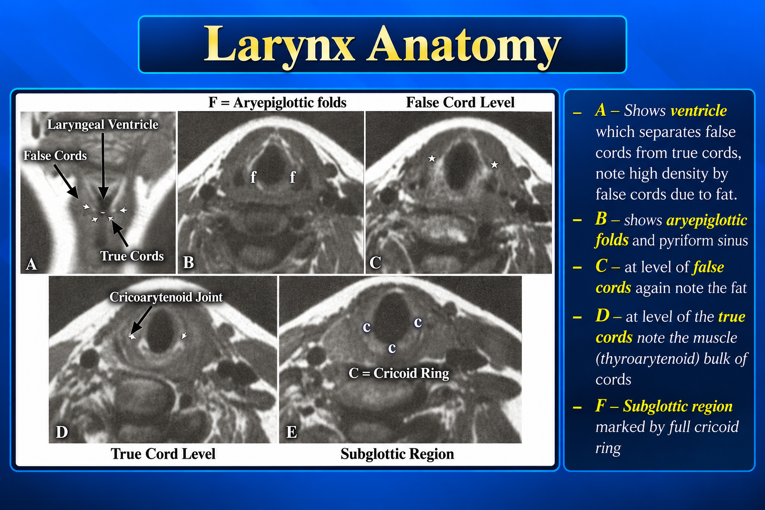
Axial laryngeal levels from aryepiglottic folds to subglottis
Axial Level-by-Level ReviewClick to enlarge
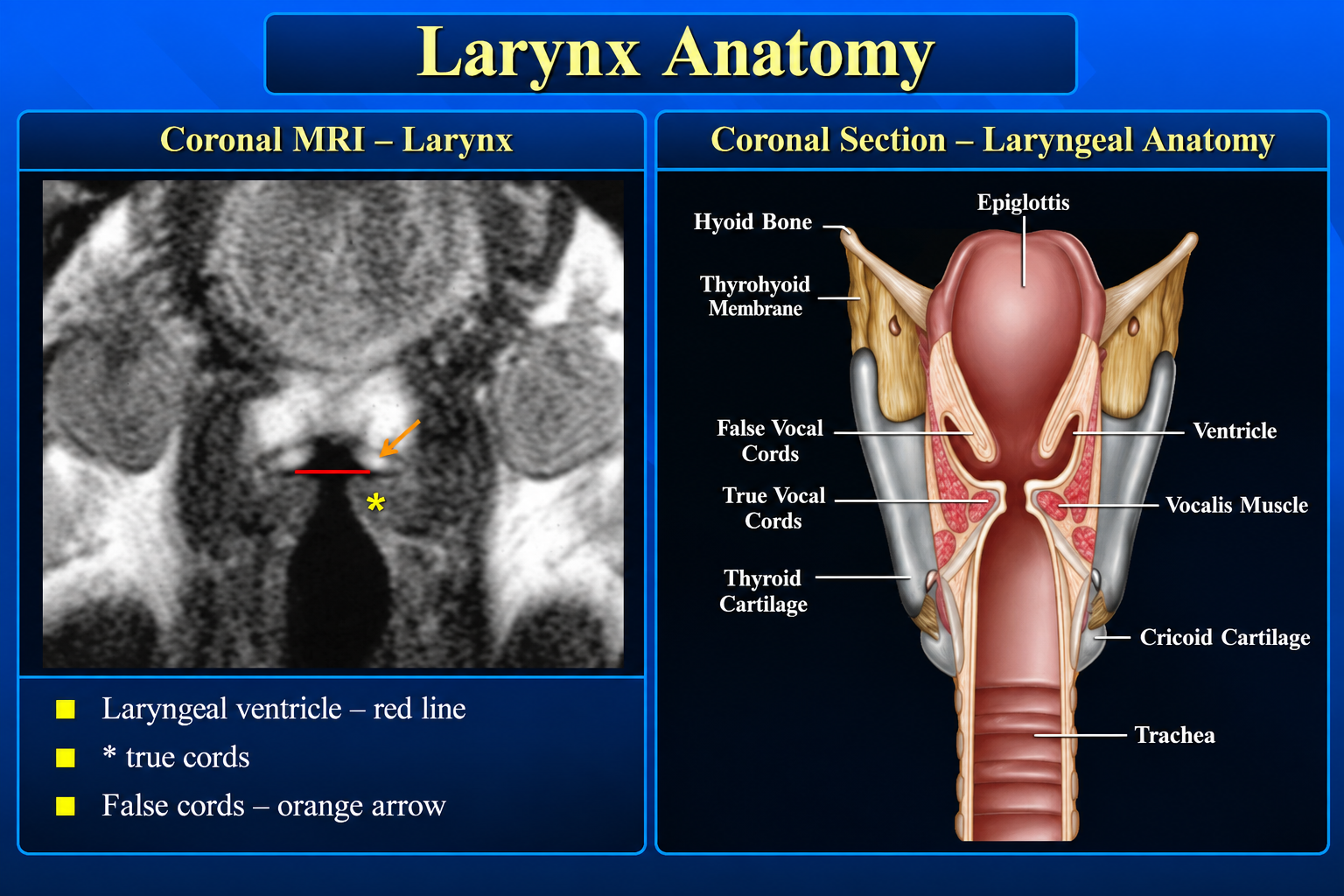
Coronal MRI and illustration of laryngeal anatomy
Coronal Laryngeal AnatomyClick to enlarge

Subsites

SupraglottisEpiglottis, aryepiglottic folds, arytenoids, false vocal cords, and laryngeal ventricles.
GlottisTrue vocal cords with the anterior and posterior commissures.
SubglottisExtends from the inferior margin of the true cords to the inferior border of the cricoid cartilage.

Cartilaginous Framework

  • Epiglottis: elastic cartilage; its free margin projects above the hyoid.
  • Thyroid: paired laminae form the anterior and lateral framework.
  • Cricoid: complete ring with a narrow anterior arch and broad posterior lamina.
  • Arytenoids: sit on the posterior cricoid and anchor the vocal processes.
  • Corniculate and cuneiform cartilages: support the aryepiglottic folds.

Deep Spaces

  • Pre-epiglottic space: fat-filled triangular space anterior to the epiglottis.
  • Paraglottic spaces: paired fat-containing spaces lateral to the laryngeal ventricle and cords.
  • The pre-epiglottic and paraglottic spaces communicate superiorly.
  • Submucosal tumor may spread through these spaces despite limited surface abnormality.
Staging relevance: Deep-space invasion can materially change local tumor stage and surgical planning.

Epiglottis and Pre-epiglottic Space

The hyoid divides the epiglottis into suprahyoid and infrahyoid portions. The suprahyoid segment forms the free upper margin; the infrahyoid segment attaches inferiorly through the thyroepiglottic ligament.

Pre-epiglottic boundaries

  • Anterior: hyoid, thyrohyoid membrane, and upper thyroid cartilage.
  • Posterior: epiglottis.
  • Superior: hyoepiglottic ligament.
  • Inferolateral: paraglottic spaces.
Vallecular landmark: The median glossoepiglottic fold separates the paired valleculae, producing the classic Mercedes-Benz configuration on axial images.

False Cords, Ventricle, and True Cords

  • False cords: supraglottic folds containing fat and minor salivary glands.
  • Ventricle: recess separating false and true cords; a useful coronal landmark.
  • True cords: paired vocal ligaments and thyroarytenoid/vocalis muscles with little normal fat.
  • Anterior commissure: anterior meeting point at Broyles ligament; normally thin and symmetric.
  • Posterior commissure: mucosa over the interarytenoid region.
Imaging pearl: The true cords are denser than the fat-containing false cords on CT. Compare cord position and symmetry for signs of fixation or paresis.

Membranes and Ligaments

StructureKey relationship
Thyrohyoid membraneBetween thyroid cartilage and hyoid; traversed by internal superior laryngeal neurovascular bundle.
Hyoepiglottic ligamentAnchors the epiglottis to the hyoid and forms the superior boundary of the pre-epiglottic space.
Thyroepiglottic ligamentAttaches the epiglottis to the inner thyroid cartilage.
Quadrangular membraneDeep to aryepiglottic fold; lower free margin forms the vestibular ligament.
Conus elasticusInferior fibroelastic membrane; upper free margin forms the vocal ligament.
Cricotracheal ligamentConnects the cricoid to the first tracheal ring.

Axial Search Pattern

LevelExpected landmarks
Suprahyoid epiglottisValleculae, median/lateral glossoepiglottic folds
HyoidPre-epiglottic fat and upper supraglottic framework
Aryepiglottic foldsLaryngeal vestibule and pyriform sinuses
False cordsFat-containing vestibular folds and paraglottic spaces
True cordsVocalis muscle, anterior commissure, arytenoids
SubglottisAirway enclosed by the complete cricoid ring

Ten-Point Imaging Checklist

  1. Airway caliber and symmetry.
  2. Epiglottic contour and thickness.
  3. Aryepiglottic folds and pyriform sinuses.
  4. False vocal cords and ventricles.
  5. True vocal cord symmetry and position.
  6. Anterior and posterior commissures.
  7. Pre-epiglottic space.
  8. Paraglottic spaces.
  9. Thyroid, cricoid, and arytenoid cartilages.
  10. Extralaryngeal spread and cervical nodes.

Suggested Reporting Language

The larynx is symmetric without focal mucosal mass. The pre-epiglottic and paraglottic fat planes are preserved. The vocal cords are symmetric in position. No cartilage destruction or extralaryngeal extension is identified.

When abnormal

State the primary subsite, cranio-caudal extent, cord mobility if known, commissural or deep-space involvement, cartilage invasion, extralaryngeal spread, and nodal disease.

Technique note

Thin-section contrast-enhanced CT is commonly used for mucosal and cartilage assessment; MRI is complementary for deep-space, marrow, and perineural evaluation.
Further Anatomy References

Secondary Links