Lymphatic Drainage
Lateral pathway
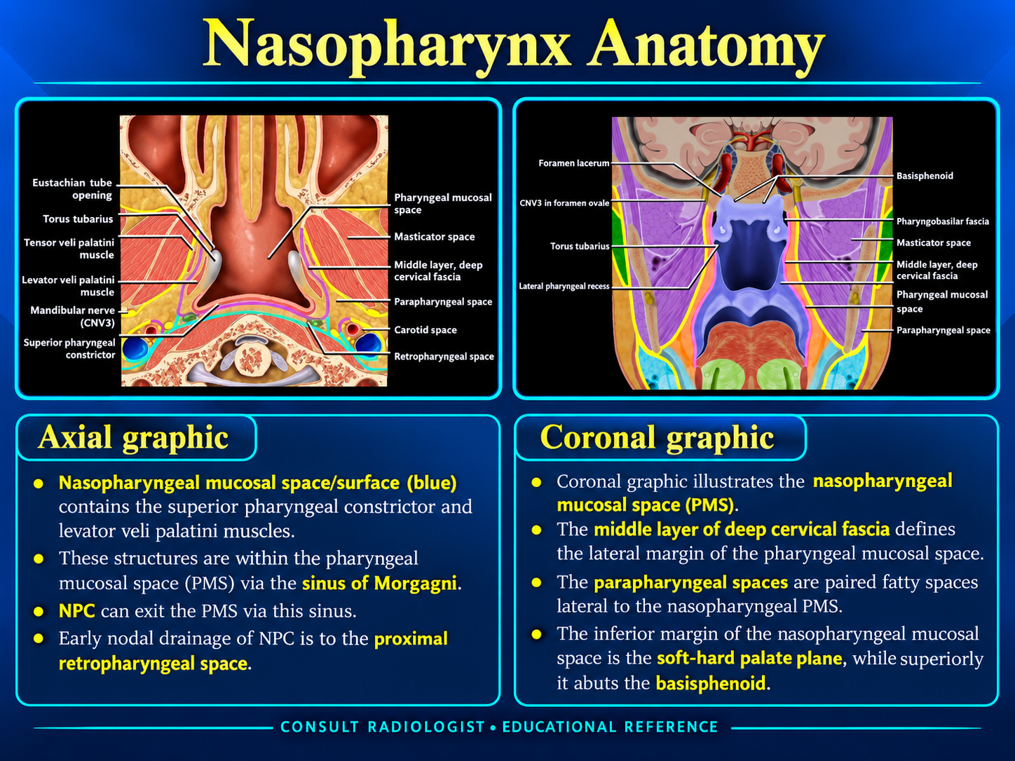
Lymphatics traverse the superior constrictor and drain to lateral retropharyngeal, upper deep cervical, and posterior-triangle nodes.
Medial pathway
The roof and posterior wall drain toward median retropharyngeal nodes.
Imaging pearl: Retropharyngeal nodes are an early and common site of spread. Nasopharyngeal carcinoma may also present with bilateral level II nodal disease.
Systemic drainage
Deep cervical nodal chains ultimately drain to the jugular lymphatic trunks.
Vessels and Innervation
Arterial supply
- Ascending pharyngeal artery.
- Ascending palatine branch of the facial artery.
- Ascending cervical artery.
- Branches of the maxillary artery.
Venous drainage
Pharyngeal and pterygoid venous plexuses drain toward the internal jugular system.
Innervation
Sensation is supplied by the pharyngeal branch of V2 and CN IX. Motor supply travels through the pharyngeal plexus, principally via CN X with contributions associated with CN IX and sympathetic fibers.
Reporting Checklist
Primary site: side, subsite, craniocaudal extent, and fossa of Rosenmüller involvement.
Local extension: nasal cavity, oropharynx, parapharyngeal space, carotid space, prevertebral muscles, clivus, sphenoid sinus, and intracranial compartment.
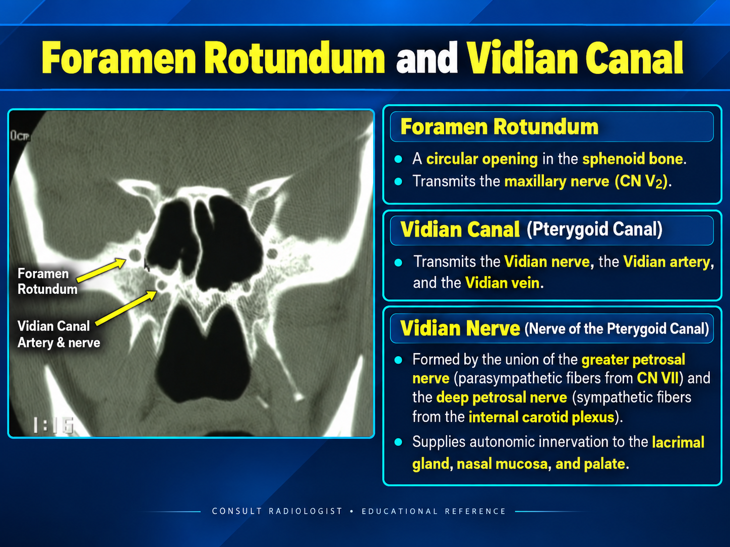
Perineural spread: V2/foramen rotundum, vidian canal, PPF, Meckel cave, and cavernous sinus.
Nodes: retropharyngeal and bilateral cervical nodal stations, including necrosis and extranodal extension.
Symmetry caveat
Normal lymphoid tissue can be prominent, especially in younger patients, but focal asymmetric mucosal thickening, deep infiltration, skull-base change, or nodal disease warrants concern.