MRI Technique and Findings
- Noncontrast axial T1 is especially sensitive to replacement of normal high-signal PPF fat.
- Acquire thin fat-suppressed postcontrast images through the deep face and skull base.
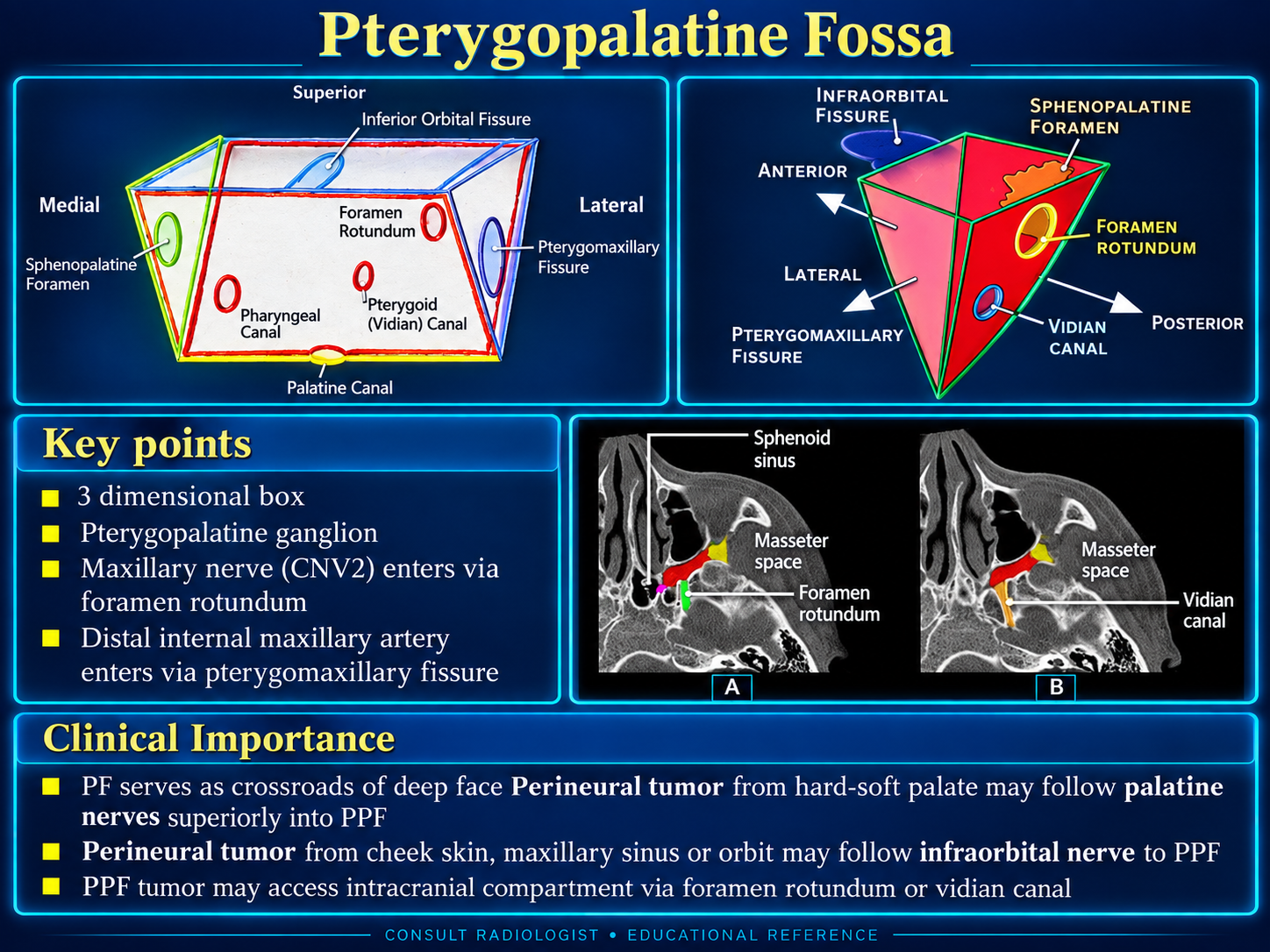
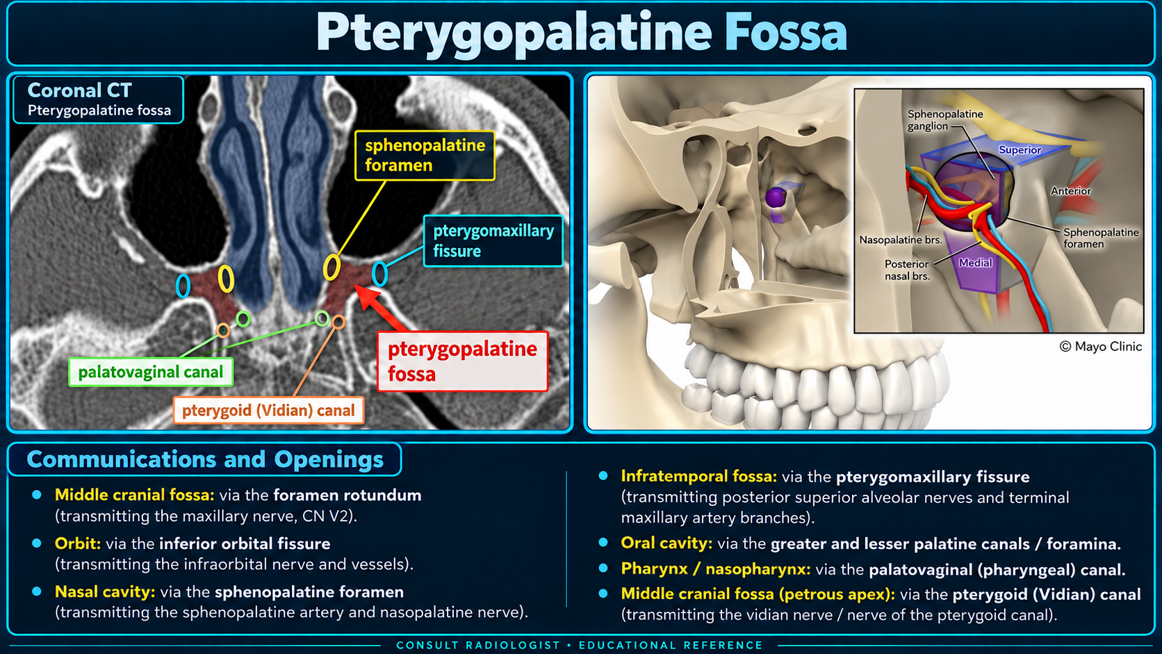
- Evaluate V2 from the PPF through foramen rotundum to Meckel cave and cavernous sinus.
- Evaluate the vidian nerve toward the foramen lacerum and the infraorbital nerve toward the orbit and cheek.
- Dental hardware and adjacent sinus air may create susceptibility artifact.
Mild symmetric enhancement may be normal; asymmetric enhancement, expansion, or loss of fat is suspicious.
Reporting Checklist
PPF: preserved or replaced fat, enhancement, mass effect, and side-to-side asymmetry.
Neural pathways: V2/foramen rotundum, vidian canal, palatine canals, infraorbital nerve, and Meckel cave/cavernous sinus.
Direct extension: nasal cavity, nasopharynx, maxillary sinus, orbit, masticator space, palate, and skull base.
Bone and vessels: foraminal enlargement, remodeling versus destruction, maxillary artery involvement, and tumor vascularity.
High-yield statement
When PPF involvement is present, explicitly state whether disease reaches the skull base or intracranial compartment and identify the most proximal extent of perineural spread.