External Eye Anatomy
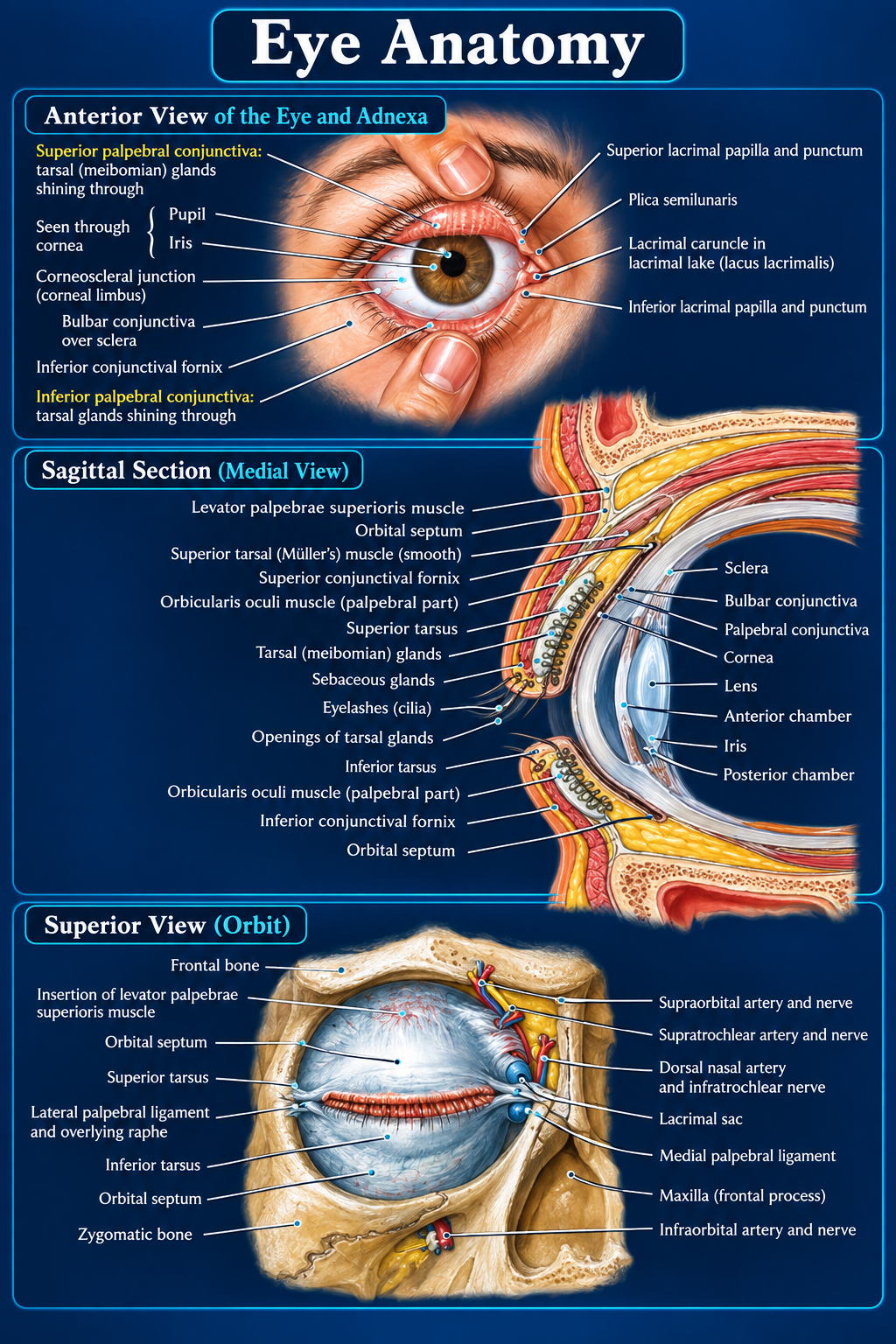
The external eye and adnexa form a mobile protective system around the anterior globe. The eyelids, conjunctiva, tarsal glands, lacrimal drainage apparatus, orbital septum, and superficial orbital neurovascular structures work together to protect and maintain the ocular surface.
Anterior Eye & Adnexa
- Pupil: central opening that admits light.
- Iris: colored diaphragm regulating pupil diameter.
- Limbus: transition between transparent cornea and opaque sclera.
- Bulbar conjunctiva: covers anterior sclera and stops at the limbus.
- Palpebral conjunctiva: lines the internal eyelid surfaces.
- Conjunctival fornices: recesses where palpebral conjunctiva reflects onto bulbar conjunctiva.
- Tarsal glands: lipid-secreting glands that stabilize the tear film and reduce evaporation.
- Lacrimal puncta: superior and inferior eyelid-margin openings that collect tears.
- Plica semilunaris: crescentic conjunctival fold at the medial canthus.
- Lacrimal caruncle: fleshy structure within the lacrimal lake.
Eyelid Support & Movement
- Levator palpebrae superioris: principal elevator of the upper eyelid.
- Superior tarsal (Müller) muscle: sympathetically innervated smooth muscle assisting lid elevation.
- Orbicularis oculi: gently closes the eyelids during blinking.
- Orbital septum: fibrous barrier separating preseptal tissues from deeper orbital contents.
- Superior and inferior tarsi: dense plates providing eyelid shape and rigidity.
- Palpebral ligaments: anchor and support the eyelids medially and laterally.
- Eyelashes: protect the ocular surface.
- Sebaceous and tarsal gland openings: deliver lipid secretions at the lid margin.
Anterior Globe
- Palpebral conjunctiva: lines the posterior eyelid.
- Bulbar conjunctiva: reflects through the fornix onto the sclera.
- Conjunctival fornices: accommodate eye movement and serve as a tear reservoir.
- Sclera: tough opaque outer coat.
- Cornea: transparent anterior refracting surface.
- Anterior chamber: aqueous-filled space between cornea and iris.
- Posterior chamber: narrow aqueous-filled space behind the iris and anterior to the lens/ciliary region.
- Lens: transparent biconvex structure providing adjustable focusing power.
Superior Orbital View
- Frontal bone: forms the orbital roof.
- Zygomatic bone: contributes to the lateral orbital rim and wall.
- Frontal process of maxilla: contributes to the inferomedial anterior rim.
- Levator insertion: joins the upper eyelid and tarsal complex.
- Orbital septum: spans the orbital aperture.
- Lacrimal sac: receives tears from the puncta and canaliculi and continues as the nasolacrimal duct.
Neurovascular Structures
- Supraorbital artery and nerve: supply and sensate the forehead and scalp.
- Supratrochlear artery and nerve: serve the medial forehead and upper eyelid.
- Dorsal nasal artery and infratrochlear nerve: serve the medial canthus, nasal root, and adjacent upper eyelid.
- Infraorbital artery and nerve: serve the lower eyelid, cheek, lateral nose, and upper lip.
Functional Integration
- Blinking by orbicularis oculi spreads the tear film across the cornea.
- Meibomian glands add the lipid layer that limits tear evaporation.
- Tears pool medially in the lacrimal lake.
- The puncta direct tears into the canaliculi and lacrimal sac.
- The nasolacrimal duct drains tears toward the nasal cavity.
- The levator and Müller muscle elevate the upper lid; the tarsi and palpebral ligaments maintain lid contour.
Lacrimal lake → puncta → canaliculi → lacrimal sac → nasolacrimal duct