Diagnostic Framework
| Pattern | More likely category | Key clues |
|---|
| Unilateral orbital-lobe mass | Epithelial neoplasm | Inferomedial globe displacement; remodeling versus destruction |
| Bilateral diffuse enlargement of both lobes | Lymphoid or inflammatory | Molds to globe; usually no destructive bone change |
| Painful, rapidly enlarging unilateral gland | Infection or malignant epithelial tumor | Inflammation/abscess versus destruction/perineural spread |
| Homogeneous restricted mass | Lymphoma | Often painless; both lobes; no destructive bone lesion |
| T2-hypointense diffuse enlargement | Fibroinflammatory / IgG4-related process | Variable enhancement; often steroid responsive |
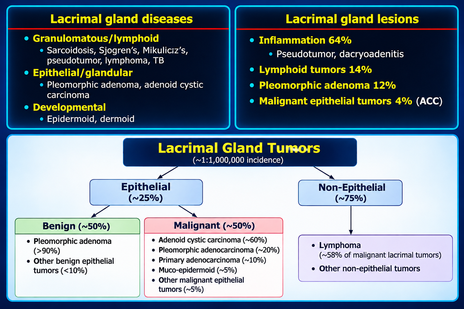
Approximate distribution: 50% inflammatory, 25% lymphoid/lymphomatous, and 25% salivary-gland–type tumors. Atypical lesions require tissue diagnosis.
Epithelial Tumors
| Lesion | Clinical pattern | Imaging | Bone / perineural behavior |
|---|
| Pleomorphic adenoma | Age 20–40; slow, painless unilateral proptosis | Well-circumscribed orbital-lobe mass; moderate enhancement | Smooth scalloping/remodeling |
| Adenoid cystic carcinoma | Often painful; sometimes paresthesia | Solid enhancing unilateral orbital-lobe mass | Destruction and marked perineural-spread propensity |
| Mucoepidermoid carcinoma | Painful, relatively rapid proptosis | Can resemble adenoid cystic carcinoma | Scalloping or destruction; local invasion |
Red flags for malignancy: pain, rapid progression, osseous destruction, and perineural extension. For suspected adenoid cystic carcinoma, inspect V1/V2 pathways, superior orbital fissure, orbital apex, cavernous sinus, foramen rotundum, and pterygopalatine fossa.