Muscles, Foramina & Nerves

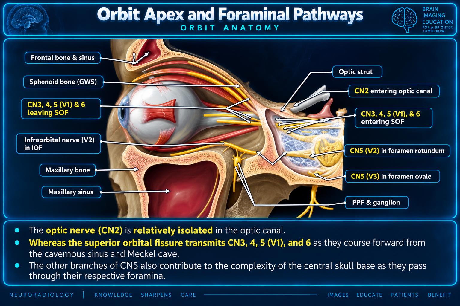
The orbital muscles, bony passages, and cranial nerves are organized around the orbital apex. The four rectus muscles arise from the annulus of Zinn, while the optic canal and orbital fissures provide distinct neurovascular pathways.Optic canal: CN II + ophthalmic artery   •   Superior orbital fissure: CN III, IV, V1, VI + superior ophthalmic vein

Reference Images

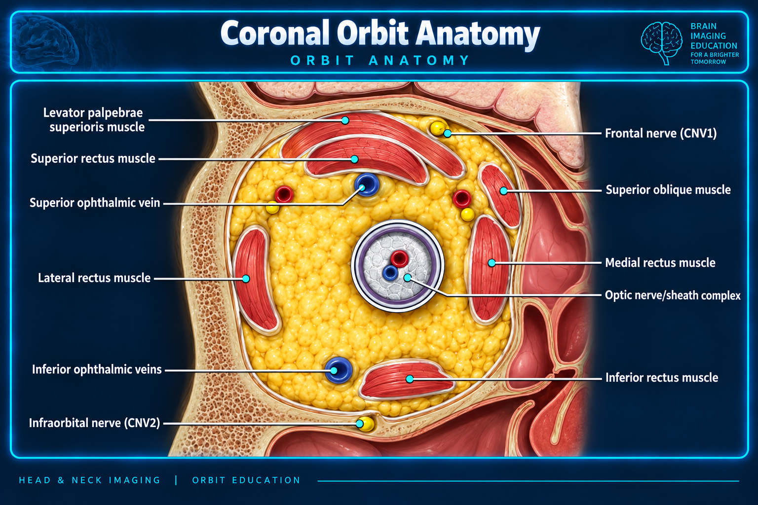
Coronal orbit anatomy
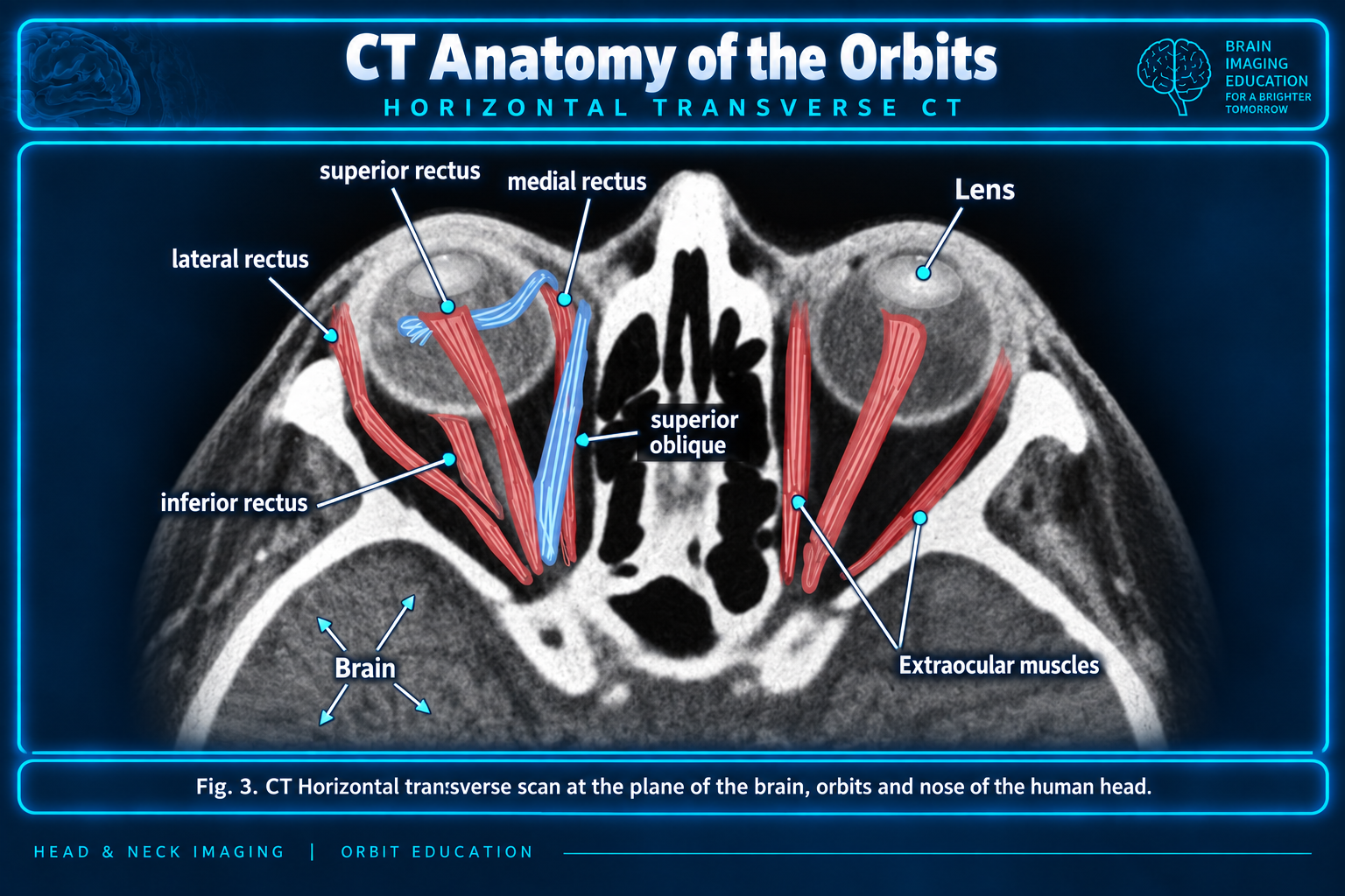
Axial CT extraocular muscle anatomy
Extraocular muscles on axial CT
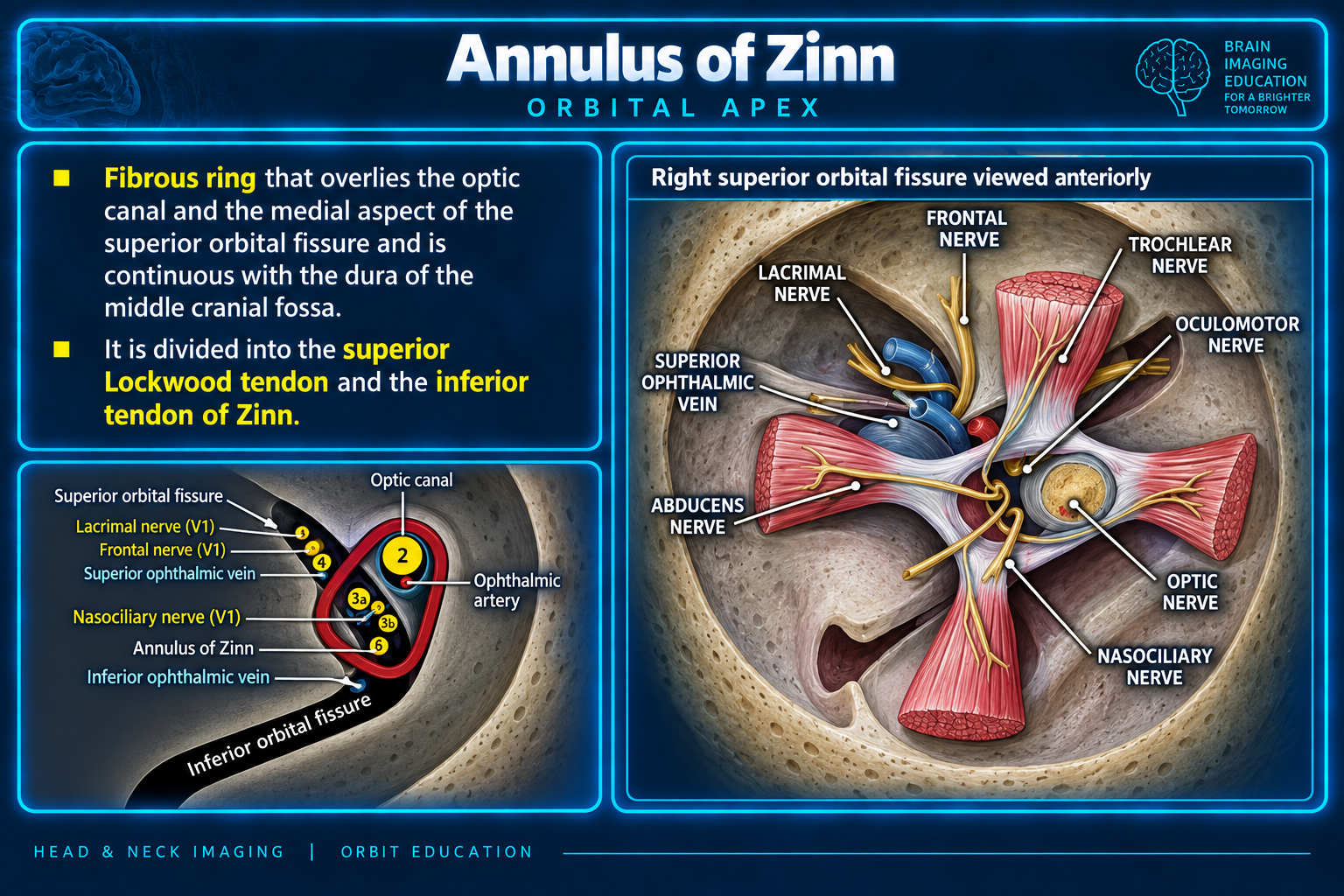
Annulus of Zinn and orbital apex
Annulus of Zinn
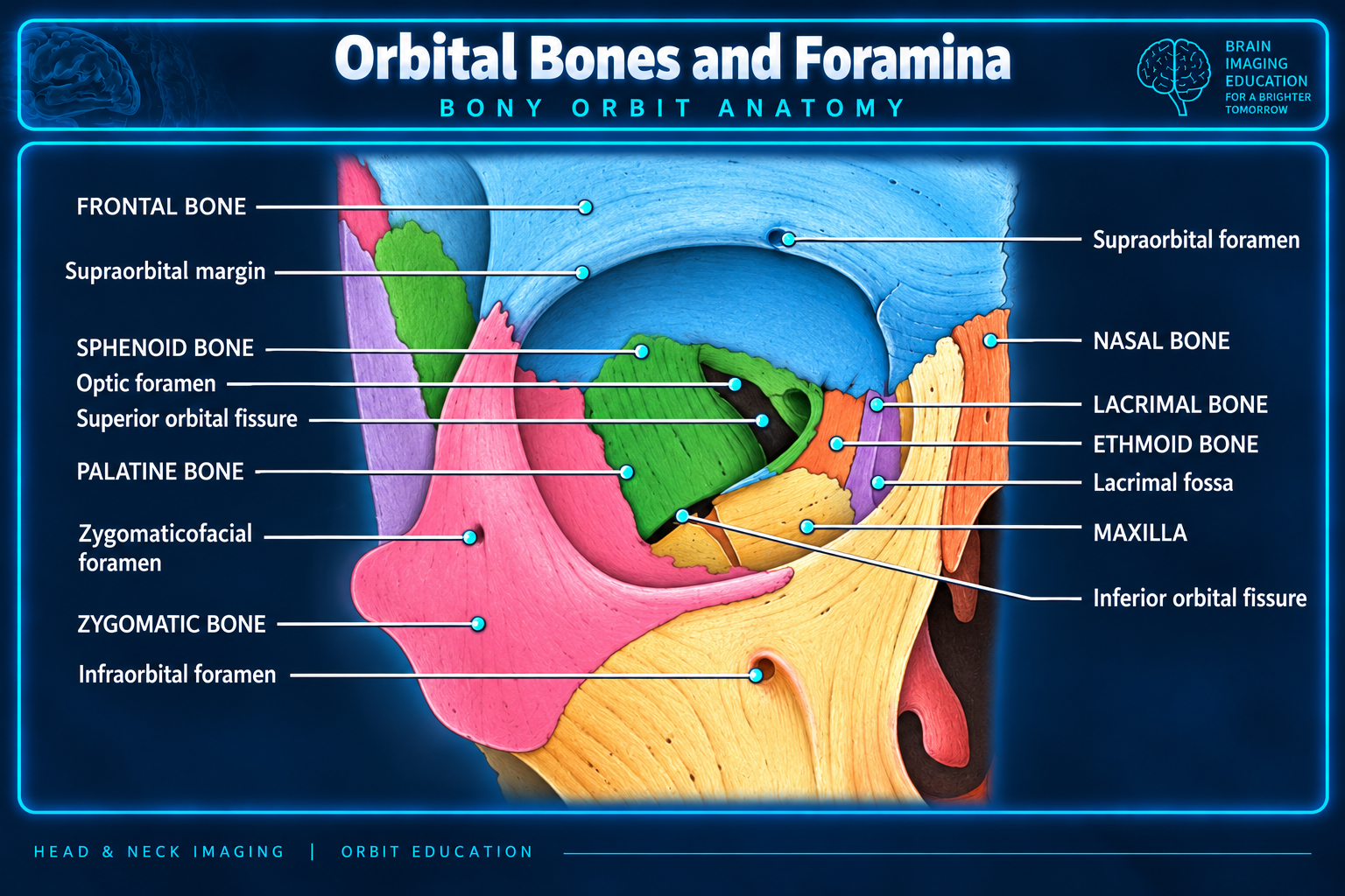
Orbital bones and foramina
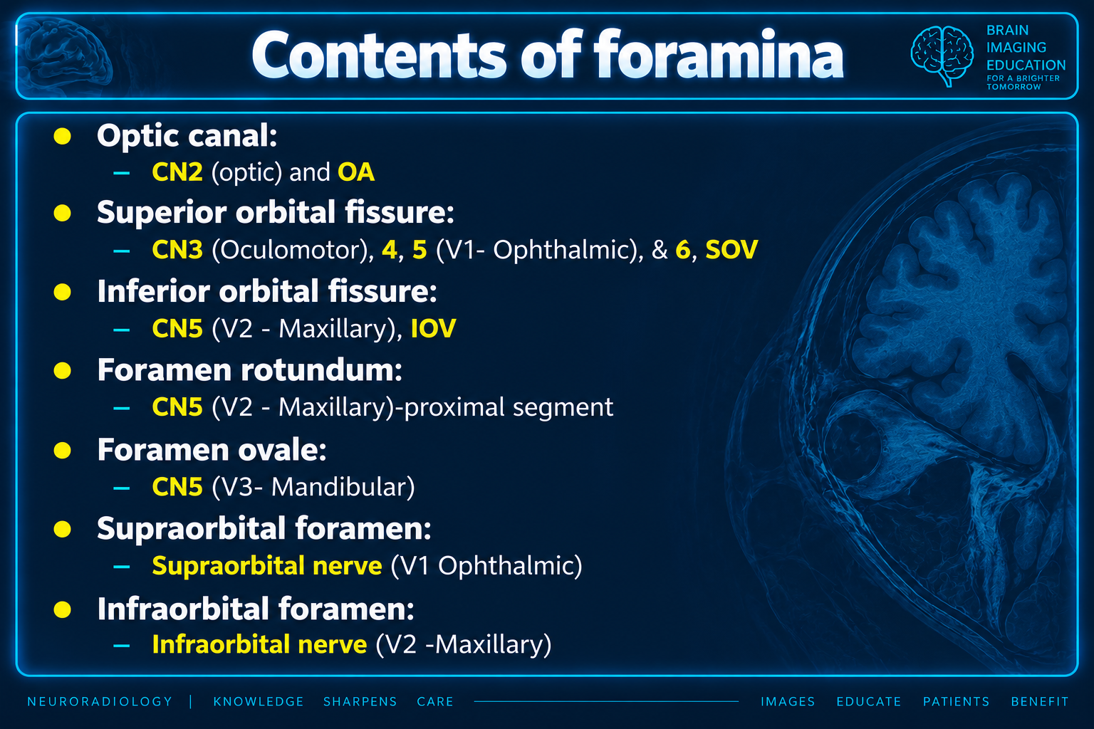
Contents of orbital and facial foramina
Contents of foramina
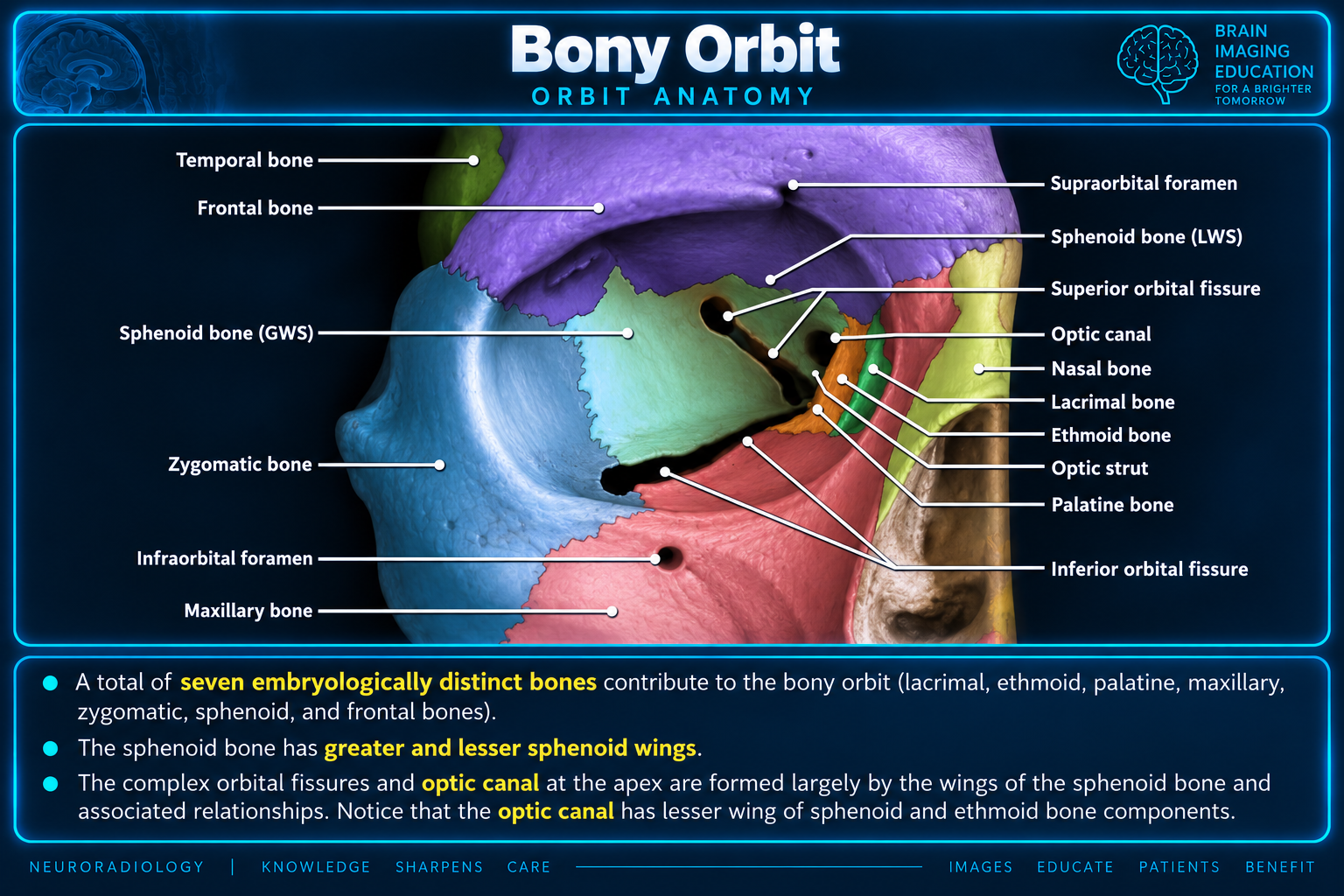
Bony orbit anatomy
Bony orbit
Orbital apex and foraminal pathways
Orbital apex pathways
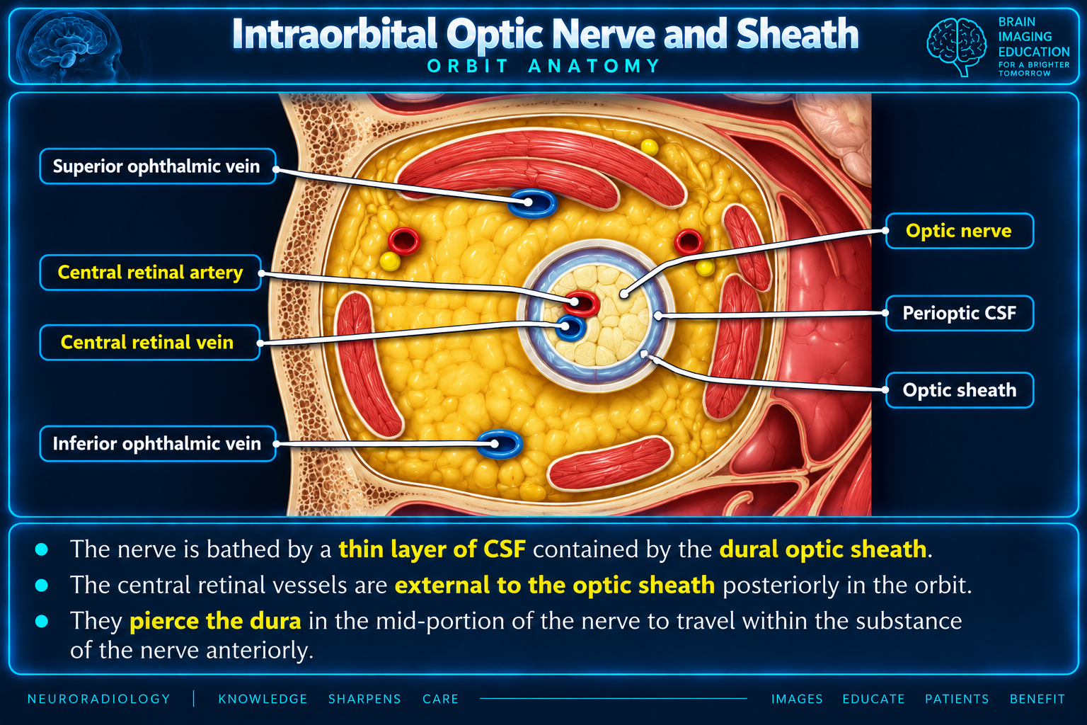
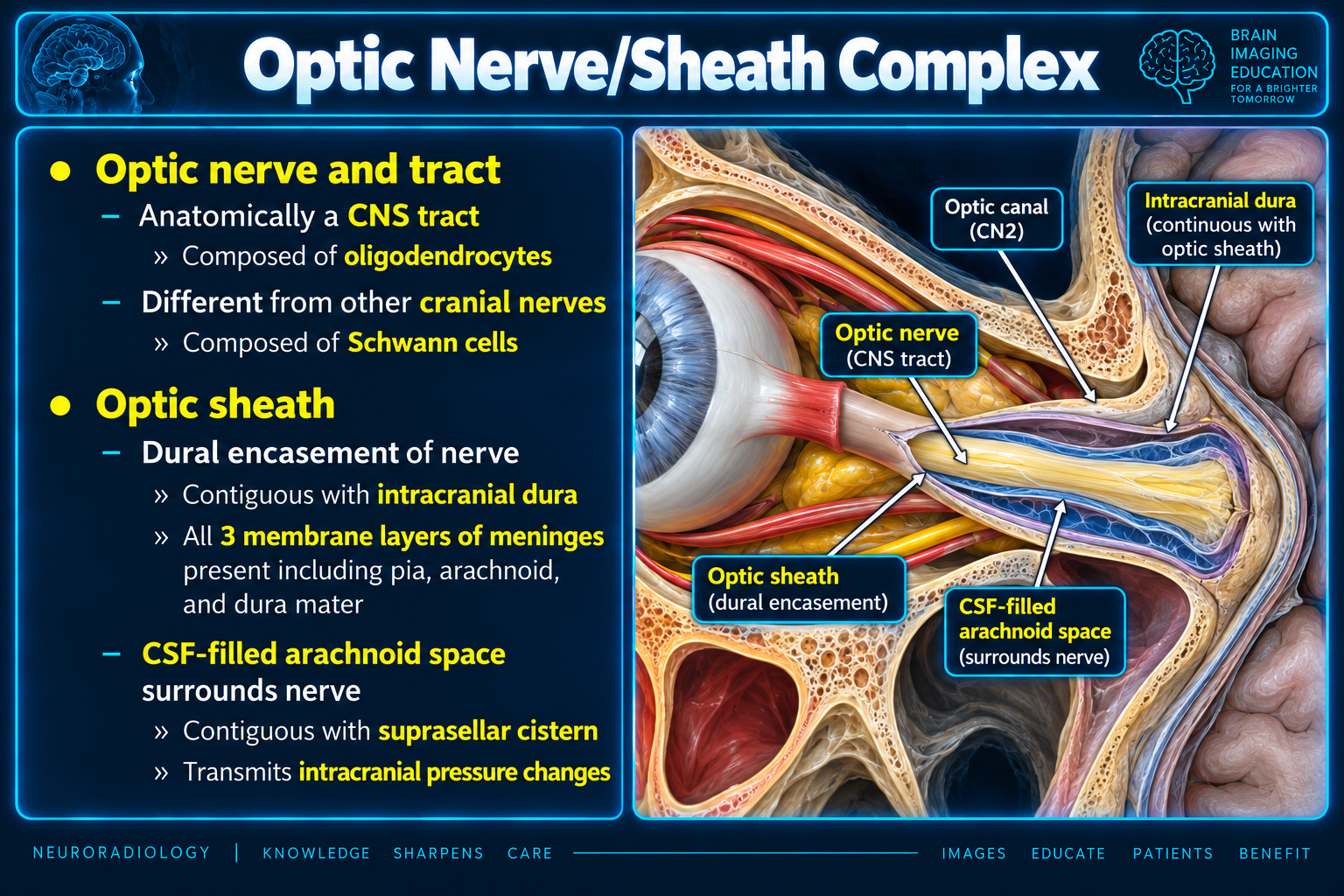
Intraorbital optic nerve and sheath
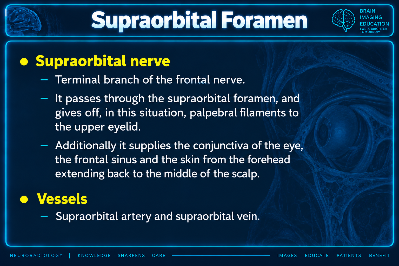
Supraorbital foramen and nerve
Supraorbital foramen
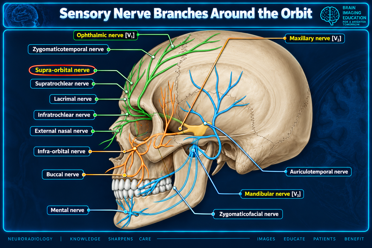
Sensory nerve branches around the orbit
Orbital sensory branches
Optic nerve and sheath complex
Optic nerve/sheath complex
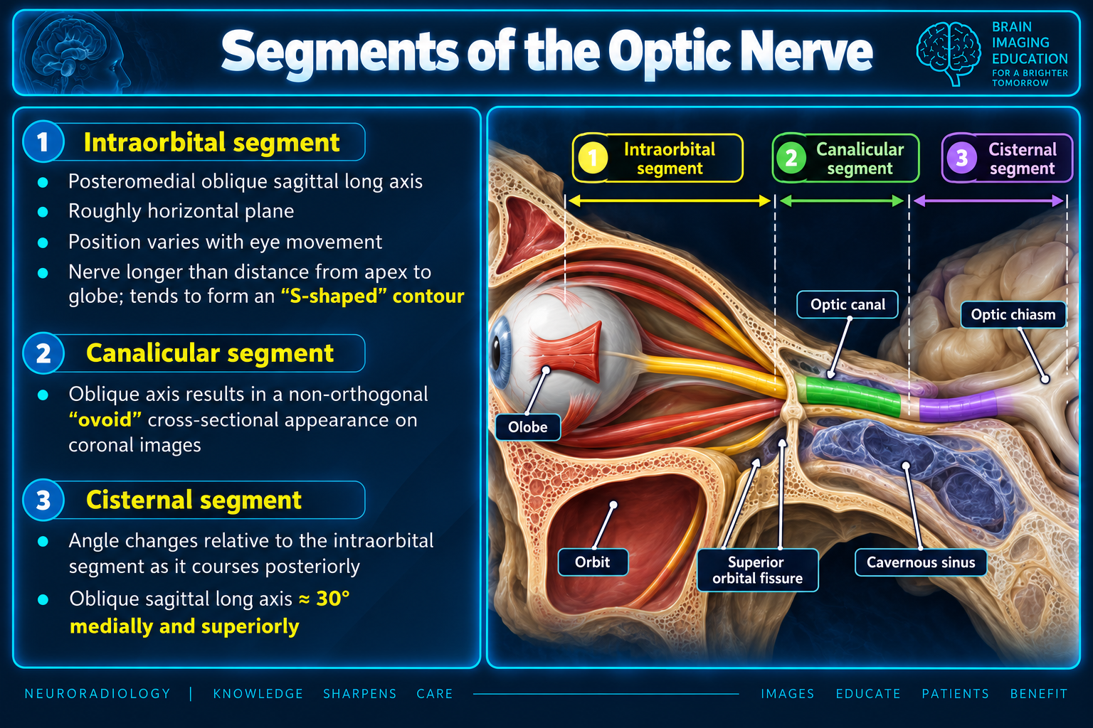
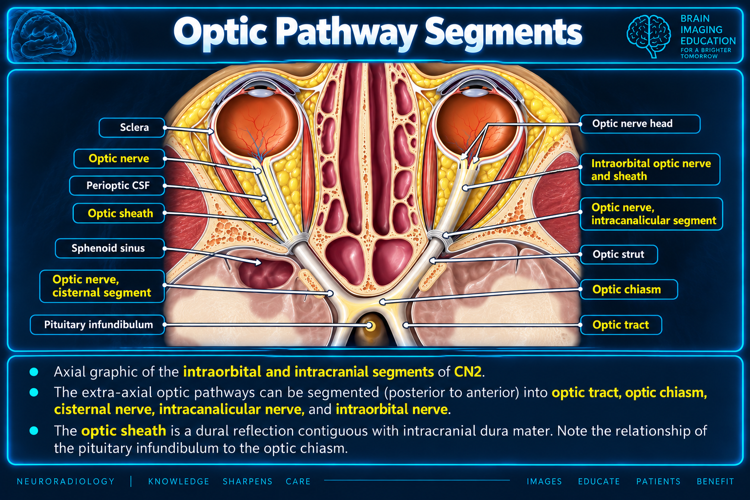
Segments of the optic nerve
Optic nerve segments
Optic pathway segments

Extraocular Muscles

MuscleOrigin / courseMain actionOther actionsNerve
Medial rectusAnnulus → medial scleraAdduction—CN III
Lateral rectusAnnulus → lateral scleraAbduction—CN VI
Superior rectusAnnulus → superior scleraElevationAdduction, intorsionCN III
Inferior rectusAnnulus → inferior scleraDepressionAdduction, extorsionCN III
Superior obliqueSphenoid → trochlea → superolateral scleraIntorsionDepression, abductionCN IV
Inferior obliqueAnterior orbital floor → inferolateral scleraExtorsionElevation, abductionCN III
LevatorLesser sphenoid wing → upper lidLid elevation—CN III

Key relationships

The recti form the muscle cone and define the intraconal space. The insertion order from closest to farthest from the limbus is medial rectus 5.5 mm → inferior rectus 6.5 mm → lateral rectus 6.9 mm → superior rectus 7.7 mm (spiral of Tillaux).

Actions & Testing

  • Mnemonic: LR6 SO4, all the rest III.
  • Superior rectus: abduct, then look up.
  • Inferior oblique: adduct, then look up.
  • Inferior rectus: abduct, then look down.
  • Superior oblique: adduct, then look down.
  • Superior oblique travels through the trochlea.
  • Inferior oblique uniquely originates in the anterior orbit.

Foramina & Fissures

OpeningConnectionPrincipal contents
Optic canalOrbital apexCN II, ophthalmic artery
Superior orbital fissureCavernous sinus region → orbitCN III, IV, V1, VI, superior ophthalmic vein
Inferior orbital fissureOrbit ↔ pterygopalatine/infratemporal regionsV2 branches, inferior ophthalmic vein
Foramen rotundumMiddle cranial fossa → PPFV2
Foramen ovaleMiddle cranial fossa → infratemporal fossaV3
Supraorbital notch/foramenSuperior orbital rimSupraorbital nerve, artery, vein
Infraorbital foramenAnterior maxillaInfraorbital nerve and vessels

Bony Orbit

  • Frontal: roof and superior rim.
  • Sphenoid: posterior orbit and apex.
  • Ethmoid: medial wall.
  • Lacrimal: anterior medial wall.
  • Maxilla: floor and inferomedial rim.
  • Zygomatic: lateral wall and rim.
  • Palatine: small posterior floor contribution.

Cranial Nerves

  • CN II: vision; passes with the ophthalmic artery through the optic canal.
  • CN III: SR, MR, IR, IO, LPS and parasympathetic pupillary fibers.
  • CN IV: superior oblique.
  • V1: orbit, cornea, conjunctiva, upper lid, forehead, and dorsum of nose.
  • V2: enters through inferior orbital fissure and continues as the infraorbital nerve.
  • CN VI: lateral rectus.

Optic Nerve & Sheath

  • The optic nerve is a CNS tract with oligodendrocytes.
  • Its dural sheath is continuous with intracranial dura and includes all three meningeal layers.
  • The perioptic subarachnoid space contains CSF and communicates with the suprasellar cistern.
  • Segments: intraorbital, canalicular, and cisternal.
  • The intraorbital segment has an S-shaped contour allowing globe movement.
  • Central retinal vessels pierce the dura in the mid-orbit and then travel within the nerve.

Palsy Patterns

  • CN III: ptosis, impaired adduction/elevation/depression, possible mydriasis; eye typically “down and out.”
  • CN IV: impaired depression in adduction; vertical/torsional diplopia worse in downgaze.
  • CN VI: impaired abduction and horizontal diplopia worse toward the affected side.

Imaging Localization

PatternSuggested localization
Visual loss without ocular-motor palsyOptic nerve or optic canal
Visual loss plus III, IV, VI palsies and V1 sensory lossOrbital apex / superior orbital fissure
III, IV, VI palsies plus V1/V2 sensory lossCavernous sinus or posterior orbital/SOF region
Lower-lid, cheek, lateral-nose, upper-lip, or upper-teeth numbnessInfraorbital nerve/V2 pathway
Forehead or scalp numbnessSupraorbital nerve / frontal V1 branch
Orbital-floor fracture with cheek paresthesiaInfraorbital canal and nerve

Muscle assessment

Report the involved muscles, tendon sparing or involvement, symmetry, adjacent fat inflammation, lacrimal-gland involvement, apical crowding, and optic-nerve compression.

Secondary Links