Nasolacrimal Anatomy & Lesions

The nasolacrimal drainage apparatus carries tears from the medial eyelids into the inferior nasal meatus. Imaging should distinguish common obstruction and inflammation from an uncommon but important obstructing neoplasm.A discrete enhancing mass, progressive unilateral symptoms, bone destruction, bloody tearing, or perineural spread should raise concern for malignancy.

Reference Images

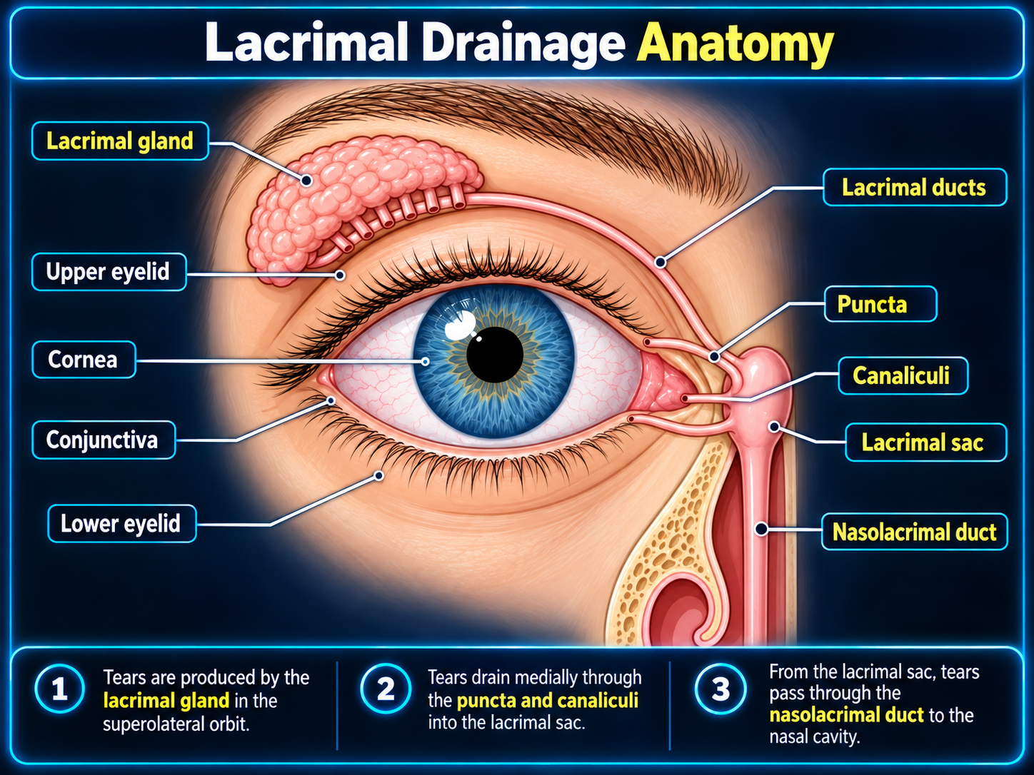
Lacrimal drainage anatomy
Lacrimal drainage anatomy and tear-flow pathway
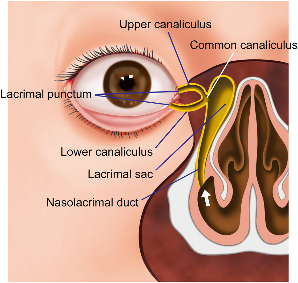
Canaliculi, lacrimal sac, and nasolacrimal duct
Puncta, canaliculi, lacrimal sac, and nasolacrimal duct
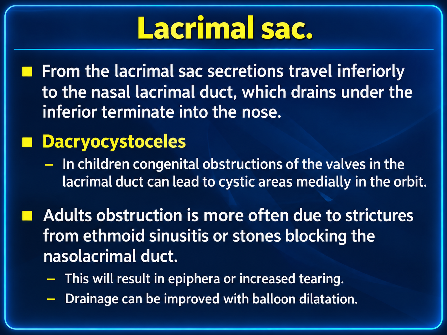
Lacrimal sac obstruction and dacryocystocele
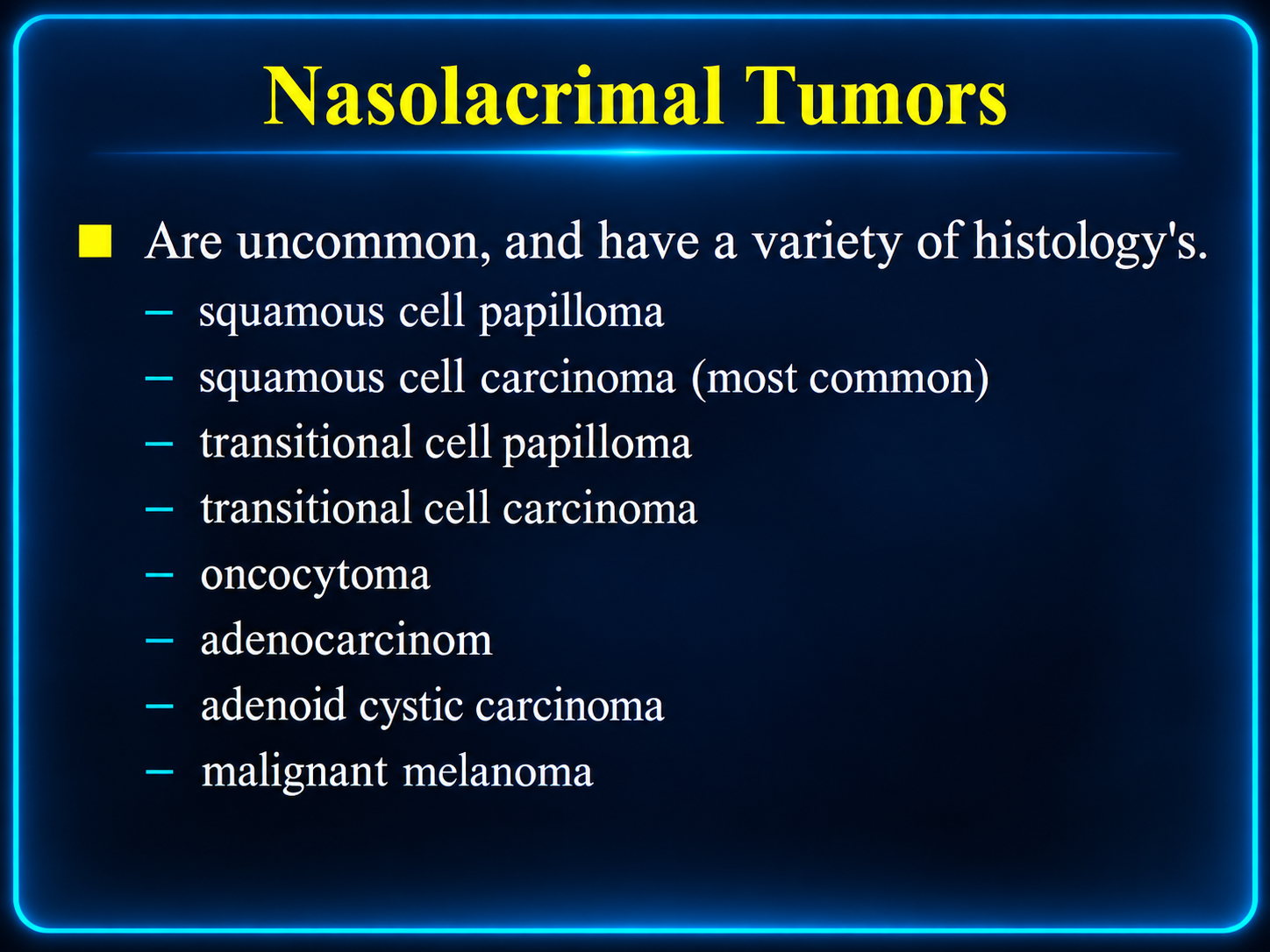
Nasolacrimal tumor histologies

Drainage Anatomy

  1. Upper and lower puncta collect tears at the medial eyelid margins.
  2. Vertical then horizontal canaliculi usually unite as a common canaliculus.
  3. The common canaliculus enters the lacrimal sac; the valve of Rosenmüller limits reflux.
  4. The sac continues as the nasolacrimal duct, approximately 18 mm long and 4.5–5 mm wide.
  5. The duct opens beneath the inferior turbinate at the inferior meatus; the valve of Hasner guards the distal opening.

The sac lies in the lacrimal fossa between the anterior and posterior lacrimal crests.

Normal Imaging & Modality Choice

ModalityBest useKey assessment
CTFirst-line structural surveyBone remodeling or destruction, calcification/dacryolith, ductal expansion, adjacent sinus disease, and abscess
MRISoft-tissue characterization and stagingEnhancement, diffusion, orbital or sinonasal extension, skull-base involvement, and perineural spread
Dacryocystography / CT dacryocystographyPrecise obstruction mappingLevel and completeness of stenosis, filling defects, fistula, and postoperative anatomy

A small amount of fluid or air may normally be present in the lacrimal sac or duct. Interpret caliber and contents with symmetry, symptoms, surrounding inflammation, and the presence or absence of an enhancing mass.

Obstruction & Inflammation

EntityTypical cause / settingImaging pattern
Congenital obstructionPersistent distal membrane at the valve of HasnerDilated sac and duct; may form a neonatal dacryocystocele
Primary acquired obstructionIdiopathic inflammation and fibrosis, typically in adultsStenosis without a focal mass; chronic sac distention may occur
Secondary acquired obstructionTrauma, surgery, sinus disease, stones, inflammation, or tumorFindings depend on cause; scrutinize the full drainage pathway
Acute dacryocystitisInfected obstructed sacEnhancing thick-walled distended sac with surrounding preseptal inflammation; abscess may develop
Chronic dacryocystitisLong-standing obstructionSac wall thickening, persistent distention, fibrosis, and recurrent inflammation
DacryocystoceleCombined proximal and distal outflow obstructionCystic medial canthal mass centered on the lacrimal sac, often with ductal dilatation
Complication check: report abscess, orbital extension, adjacent sinus disease, and any intracranial spread.

Obstruction Categories

  • Congenital: most often distal membranous obstruction.
  • Primary acquired: idiopathic stenosis from chronic inflammation and fibrosis.
  • Secondary acquired: infectious, inflammatory, traumatic, iatrogenic, lithiasis-related, or neoplastic.
  • Functional: impaired lacrimal pump despite an anatomically patent pathway.

Clinical consequences

  • Epiphora and recurrent discharge
  • Medial canthal swelling or tenderness
  • Recurrent dacryocystitis
  • Rarely, bloody tears or a firm painless mass

Tumors & Mimics

LesionImaging / clinical clues
Squamous cell carcinomaMost common malignant epithelial tumor; enhancing obstructing mass with possible bone destruction and local invasion
Transitional cell carcinomaArises from Schneiderian-type epithelium; may extend along the sac and duct
LymphomaRelatively homogeneous soft-tissue mass, often with restricted diffusion and less early bone destruction
Adenocarcinoma / adenoid cystic carcinomaInfiltrative enhancing mass; adenoid cystic carcinoma warrants careful evaluation for perineural spread
MelanomaMay show intrinsic T1 hyperintensity and susceptibility depending on melanin or hemorrhage
Metastasis or direct invasionConsider spread from adjacent sinonasal, cutaneous, orbital, or systemic malignancy
Benign mimicsPapilloma, granulomatous disease, inflammatory tissue, dacryolith, foreign body, mucocele, and dacryocystocele

Malignancy Red Flags

  • Progressive unilateral epiphora
  • Bloody tears or recurrent unexplained bleeding
  • Firm, painless medial canthal mass
  • Mass extending above the medial canthal tendon
  • Discrete enhancing soft tissue rather than simple fluid distention
  • Irregular bone erosion or destruction
  • Orbital, sinonasal, skin, skull-base, or intracranial invasion
  • Perineural spread or suspicious regional nodes

Reporting Checklist

  1. Epicenter: punctum/canaliculus, lacrimal sac, nasolacrimal duct, orbit, or sinonasal cavity.
  2. Morphology: fluid distention, wall thickening, filling defect, or solid enhancing mass.
  3. Extent: cranio-caudal length and involvement of the sac, duct, inferior meatus, and adjacent soft tissues.
  4. Bone: smooth remodeling, canal widening, pressure erosion, or aggressive destruction.
  5. Inflammation: preseptal/orbital stranding, collection, abscess, or fistula.
  6. Tumor staging: orbital, sinonasal, cutaneous, skull-base, intracranial, and perineural extension.
  7. Nodes and cause: regional adenopathy, sinus disease, dacryolith/foreign body, trauma, or prior surgery.

Practical Impression

State whether the appearance favors simple obstruction, active dacryocystitis, dacryocystocele, or an obstructing mass.

If a mass is present, identify its epicenter and full extent, describe bone response, and recommend contrast-enhanced MRI when soft-tissue or perineural staging is incomplete.

Do not dismiss: unilateral recurrent “dacryocystitis” with a solid enhancing component should be treated as a possible neoplasm until proven otherwise.

Secondary Links