Mandibular osteoradionecrosis
Key Images
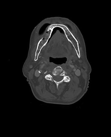
![7-year-old male patient who underwent radiation for left-sided poorly differentiated squamous cell carcinoma of the nasopharynx.
A, Axial CT scan (bone window) shows ORN involving the mandibular ramus contralateral to the primary tumor site with cortical disruption and trabecular disorganization.
B, Unenhanced T1-weighted (400/9 [TR/TE]) MR image shows abnormal homogeneous signal hypointensity of the bone marrow of the right mandible (asterisk) and cortical disruption (arrowheads). Note that the right masticator muscles appear slightly larger than the contralateral side.
C, Axial T2-weighted (4000/105 [TR/TEeff]) MR image shows abnormal hyperintense signal of the marrow (asterisk) and adjacent masseter and pterygoid muscles (arrows).
D, Contrast-enhanced fat-saturated T1-weighted (400/9 [TR/TE]) MR image shows diffuse intense enhancement of the marrow (asterisk) and adjacent musculature (arrows).](assets/image-10.png)
Look For First
- Bony sclerosis—nearly pathognomonic for ORN; absent in recurrent tumor
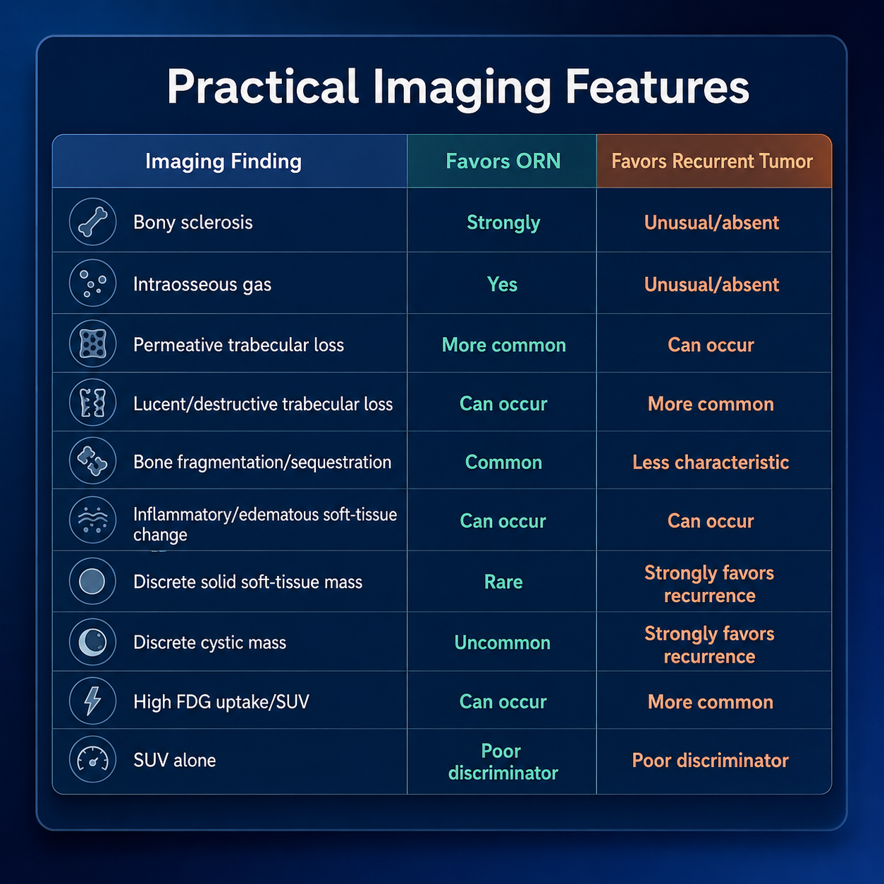
- Presence or absence of discrete soft-tissue mass—solid or cystic mass strongly suggests recurrence rather than ORN
- Pattern of trabecular bone loss: permeative loss favors ORN; lucent destructive loss favors recurrence
- Intraosseous gas, suggesting superimposed osteomyelitis in ORN
Key Image Findings
- CT is the primary imaging modality with 90% reported accuracy; CBCT provides high-detail osseous assessment with lower radiation dose than conventional CT.
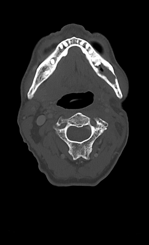
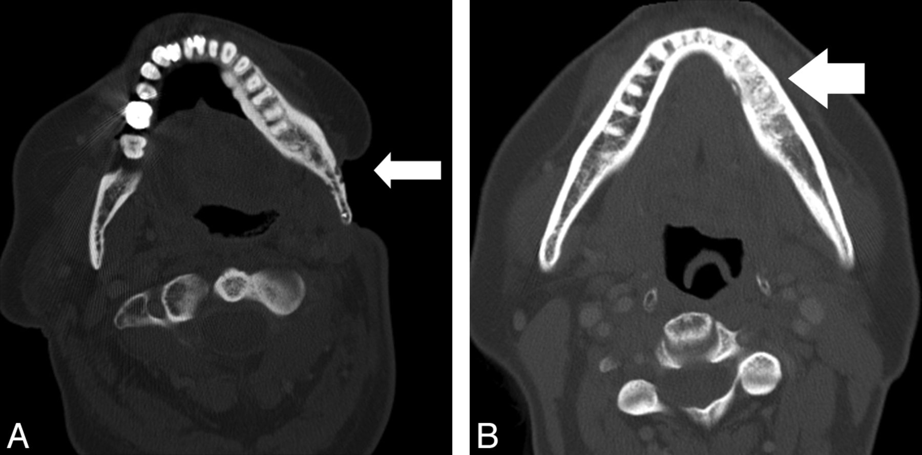
- Classic CT findings of ORN include cortical disruption or destruction, trabecular loss, bone fragmentation, bony sequestration (particularly buccal cortex), mixed sclerotic-lucent osseous change, and intraosseous gas.
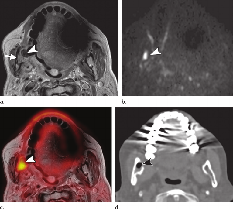
- Bony sclerosis is present in approximately one-third of ORN patients but was absent in all recurrent tumor cases in one comparative study, making it a strongly favoring feature when present.
- Permeative pattern of trabecular loss is significantly more common in ORN and reflects the hypoxic, hypocellular, hypovascular bone with impaired remodeling ability from radiation injury.
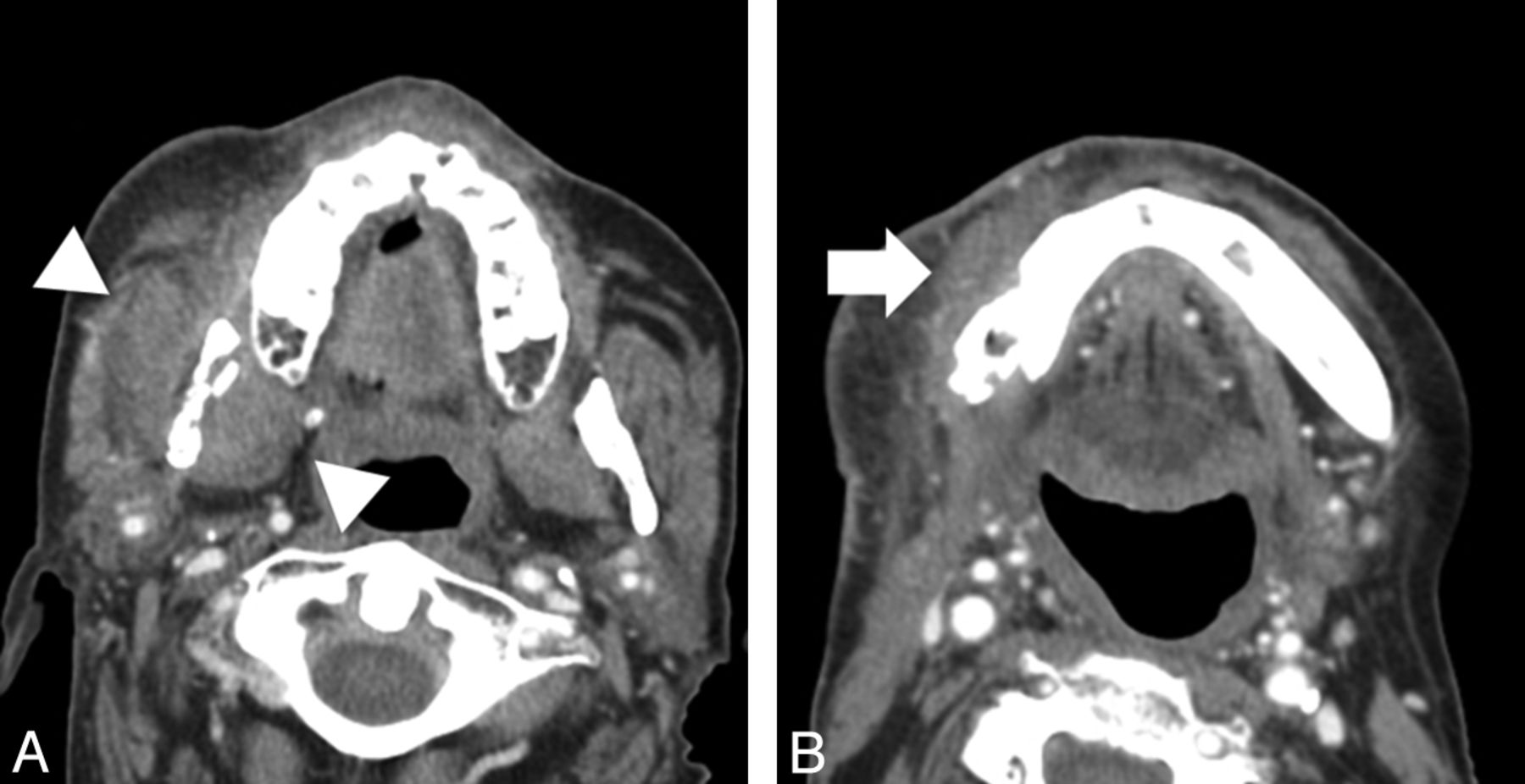
- Discrete asymmetric solid soft-tissue mass is present in more than half of recurrent malignancy but only ~2% of ORN, making this finding strongly suggestive of recurrence.
- Discrete cystic mass is present in nearly half of recurrent tumor patients but fewer than 10% of ORN; cystic lesions adjacent to abnormal bone should not automatically be attributed to abscess but must raise concern for neoplasm.
- MRI demonstrates 92% specificity for soft-tissue and vascular evaluation and is preferred when detailed soft-tissue assessment is needed, particularly for suspected recurrence.
- PET/CT demonstrates higher SUV values in recurrent tumors but with substantial overlap to ORN, making SUV measurements unreliable as isolated discriminators in individual patients.
Differential Diagnosis
- Recurrent malignancy—distinguished by presence of discrete soft-tissue mass and lucent destructive bone loss pattern; higher SUV values but substantial overlap with ORN
- Medication-related osteonecrosis of the jaw (MRONJ)—different etiology related to antiresorptive medications rather than radiation
- Chronic osteomyelitis—may demonstrate intraosseous gas but typically associated with acute clinical presentation; distinguish from ORN by clinical history
- Radiation-induced secondary neoplasia—occurs as late complication of radiation therapy; distinguished by presence of aggressive tumor mass rather than osteoradionecrosis pattern
Discussion
Mandibular ORN is more common than other forms of radiation osteonecrosis due to superficial position of mandible exposing it to relatively high radiation doses.
ORN is defined as exposed devitalized bone persisting >3 months in a patient without active or recurrent neoplastic disease, with incidence approximately 20% (range 5-37%).
Risk factors include radiation dose >60 Gy, chemotherapy with cisplatin, dental extractions (before or after radiation), poor oral hygiene, and diabetes.
Pathophysiology involves radiation-induced hypoxic, hypocellular, and hypovascular tissue environment with impaired bone turnover and remodeling capacity.
Morphologic imaging findings (particularly bony sclerosis, permeative trabecular loss, and presence/absence of soft-tissue mass) are considerably more useful than SUV measurements for distinguishing ORN from recurrence.
Plain radiography has limited utility and frequently fails to identify early disease; CT is recommended for diagnosis with reported 90% accuracy.
Reporting Pearls
When describing mandibular bone changes in a post-radiation patient, explicitly comment on: (1) presence or absence of discrete soft-tissue mass (critical discriminator—solid or cystic mass favors recurrence), (2) pattern of trabecular loss (permeative vs. lucent/destructive), and (3) presence of sclerosis (strongly favors ORN if present). State that conventional morphologic CT findings are more reliable than SUV measurements for distinguishing ORN from recurrence.
Pitfalls
- False attribution of cystic lesions to abscess or osteomyelitis without considering recurrent neoplasm; approximately 50% of recurrent tumors present with cystic mass component compared to <10% of ORN.
- Relying on SUV measurements as isolated discriminators despite substantial overlap between ORN and recurrent tumor; markedly elevated SUV increases concern but does not exclude ORN.
- Overinterpreting nonspecific inflammatory and edematous soft-tissue changes as indicative of recurrence; ORN can produce significant surrounding soft-tissue abnormality without discrete mass, which favors ORN rather than recurrence.
- Underappreciating bony sclerosis as a strongly favoring finding for ORN when present; its presence in the absence of a soft-tissue mass should strongly favor osteoradionecrosis over recurrence.