Overview
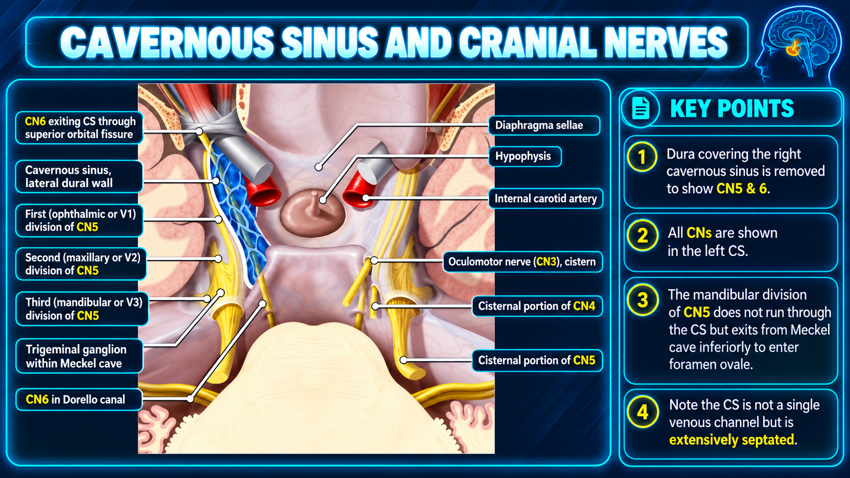
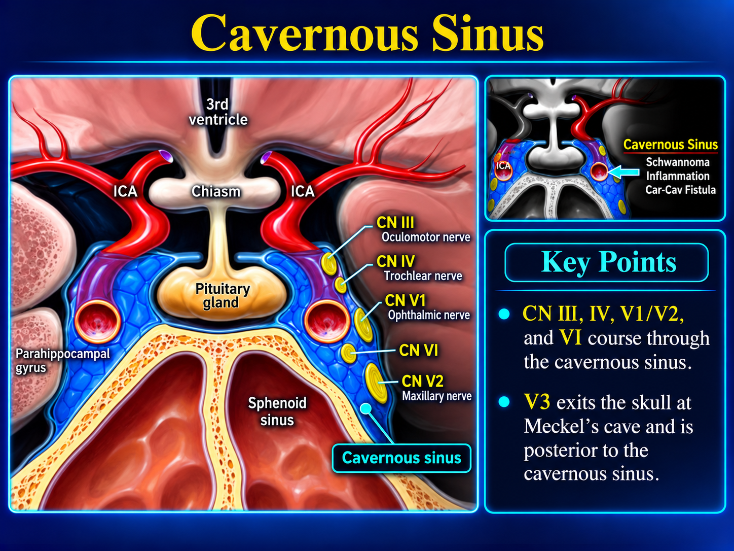
The cavernous sinuses are paired dural venous sinuses on either side of the sella turcica. They drain facial and cerebral veins and contain the internal carotid artery, cranial nerves III, IV, and VI, and the ophthalmic and maxillary divisions of cranial nerve V.
Normal contourThe normal lateral wall should be straight or concave. Convexity, asymmetry, abnormal enhancement, or expansion should prompt evaluation for pathology.
Gross Anatomy
The cavernous sinus lies on either side of the pituitary fossa and sphenoid body, between the endosteal and meningeal dural layers. It extends from the orbital apex to the petrous apex.
- Numerous fibrous septa divide it into small venous spaces.
- The pituitary gland lies medially.
- The internal carotid artery and CN VI traverse the venous compartment.
- CN III, IV, V1, and V2 course in the lateral wall.
Boundaries
- Roof: Dural fold attached to the anterior and middle clinoid processes.
- Anterior: Medial end of the superior orbital fissure.
- Posterior: Petrous apex.
- Medial: Endosteum over the sphenoid body.
- Lateral: Dura extending from the roof ridge to the middle cranial fossa floor.
- Floor: Endosteum over the base of the greater sphenoid wing.
Relations
- Superior: Middle cerebral artery, optic chiasm, and temporal uncus.
- Anterior: Orbital apex.
- Posterior: Cerebral peduncle.
- Medial: Pituitary fossa and pituitary gland.
- Lateral: Medial temporal lobe and posteroinferior Meckel cave.
- Inferior: Sphenoid sinus.
Dural Anatomy
The lateral wall is primarily a continuation of the meningeal dura from the middle cranial fossa. It passes over the cavernous sinus to the clinoid processes and diaphragma sellae, then descends around the pituitary gland to form the medial wall. The floor is formed by the endosteal layer covering the sphenoid bone. The intercavernous sinus separates this layer from the meningeal layer beneath the pituitary.
The cranial nerves in the lateral wall are enclosed by thin connective tissue. This produces a thick outer meningeal layer and a thinner translucent inner layer surrounding the nerves.
Venous Inflow
- Superior and inferior ophthalmic veins.
- Intercavernous sinus.
- Sphenoparietal sinus.
- Superficial middle cerebral vein.
- Occasionally the central retinal vein or a frontal tributary of the middle meningeal vein.
Depending on pressure gradients, the superior ophthalmic vein may drain toward or away from the cavernous sinus.
Venous Drainage
- Superior petrosal sinus toward the transverse sinus.
- Inferior petrosal sinus directly to the jugular bulb.
- Pericarotid venous plexus to the clival or basilar plexus.
- Emissary veins through the foramina Vesalii, ovale, and lacerum.
- Foramen ovale communicates with the pterygoid venous plexus.
- Paired cavernous sinuses communicate through intercavernous sinuses.
Cranial Nerves
The arrangement can be recalled with the mnemonic O TOM CAT.
Lateral wall, superior to inferior
- CN III — oculomotor nerve.
- CN IV — trochlear nerve.
- CN V1 — ophthalmic division.
- CN V2 — maxillary division, at the inferolateral wall.
Traversing the sinus
- CN VI — abducens nerve, inferolateral to the internal carotid artery.
Artery and Sympathetics
The internal carotid artery enters the posteroinferior sinus and curves as the cavernous carotid siphon, corresponding to the C4 segment.
- Meningohypophyseal trunk arises from this segment.
- Inferolateral trunk also arises here.
- A sympathetic plexus from the superior cervical ganglion surrounds the artery.
Imaging and Clinical Pearls
- Evaluate lateral-wall contour, symmetry, enhancement, and cavernous ICA caliber.
- CN VI is particularly vulnerable because it courses freely beside the ICA rather than within the lateral wall.
- Processes may extend anteriorly to the orbital apex or posteriorly toward Meckel cave and the petrous apex.
- Facial and orbital venous communications permit spread of infection and reversal of ophthalmic venous flow.
- Fat may be present within the cavernous sinus, especially with obesity or corticosteroid therapy.
References