Overview
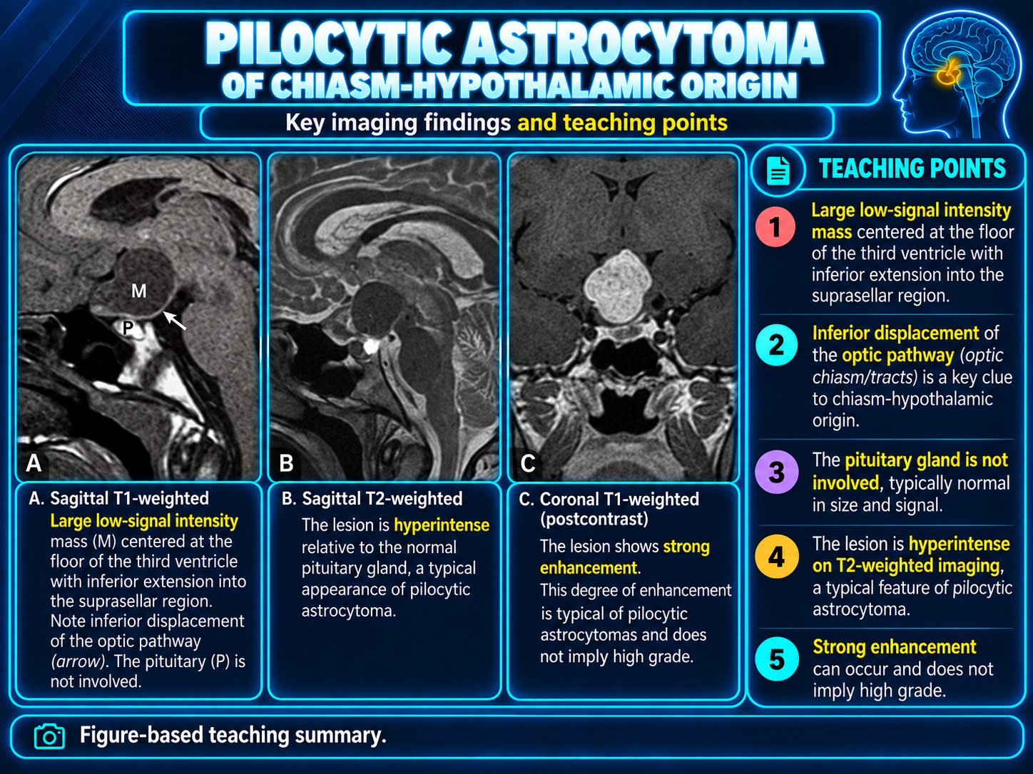
Hypothalamic astrocytoma usually refers to a pediatric optic pathway-hypothalamic glioma, most often pilocytic astrocytoma, CNS WHO grade 1. These tumors commonly form a continuum involving the optic nerves, chiasm, tracts, hypothalamus, and floor of the third ventricle rather than remaining confined to the hypothalamus.
Highest-yield localization clueDemonstrate continuity with an expanded optic pathway. Enhancement intensity is variable and is less diagnostically useful than the tumor's longitudinal anatomic distribution.
Clinical Setting
- Visual loss, optic atrophy, nystagmus, or strabismus.
- Endocrine dysfunction, growth disturbance, precocious puberty, or obesity.
- Hydrocephalus or diencephalic syndrome in infants.
- Assess NF1 status because NF1-associated tumors may differ in morphology and behavior.
- Visual and endocrine findings may progress despite stable enhancement.
Morphology and Spread
- Well-marginated, lobulated, oval, or rounded mass centered on the chiasm-hypothalamus.
- May expand the optic tracts, project into the suprasellar cistern, or extend into the third ventricle.
- Can grow around the infundibulum without originating from it.
- Large lesions may be heterogeneous or solid-cystic.
- Optic nerves may be fusiform, elongated, kinked, or buckled.
Pilocytic Pattern
Pilocytic astrocytoma is a circumscribed astrocytic glioma, CNS WHO grade 1. Optic pathway-hypothalamic tumors tend to be more solid and extend along visual pathways rather than showing the classic cerebellar cyst-with-mural-nodule pattern.
- T1 hypo- or isointense.
- T2/FLAIR hyperintense.
- Usually facilitated diffusion.
- Enhancement ranges from absent to intense and does not establish grade.
Typical MRI Appearance
| Sequence | Typical low-grade appearance | Concerning deviation |
|---|
| T1 | Isointense to hypointense to gray matter | Intrinsic T1 hyperintensity suggests hemorrhage, proteinaceous material, fat, or another lesion type |
| T2/FLAIR | Hyperintense; larger lesions may become heterogeneous | Marked T2 hypointensity suggests a hypercellular lesion such as germinoma or lymphoma |
| Postcontrast T1 | Absent, mild, homogeneous, heterogeneous, peripheral, or nodular enhancement | Thick irregular enhancement, necrosis, and infiltrative edema raise concern for high-grade tumor |
| DWI/ADC | No true restriction with increased diffusivity | Low ADC raises germinoma, lymphoma, or higher-grade neoplasm |
| SWI/GRE | Usually no blood products | Hemorrhage may occur, especially with pilomyxoid-pattern tumors |
| Perfusion | Often not markedly elevated | Elevated relative cerebral blood volume supports a vascular or higher-grade lesion but is not diagnostic |
Pilomyxoid Pattern
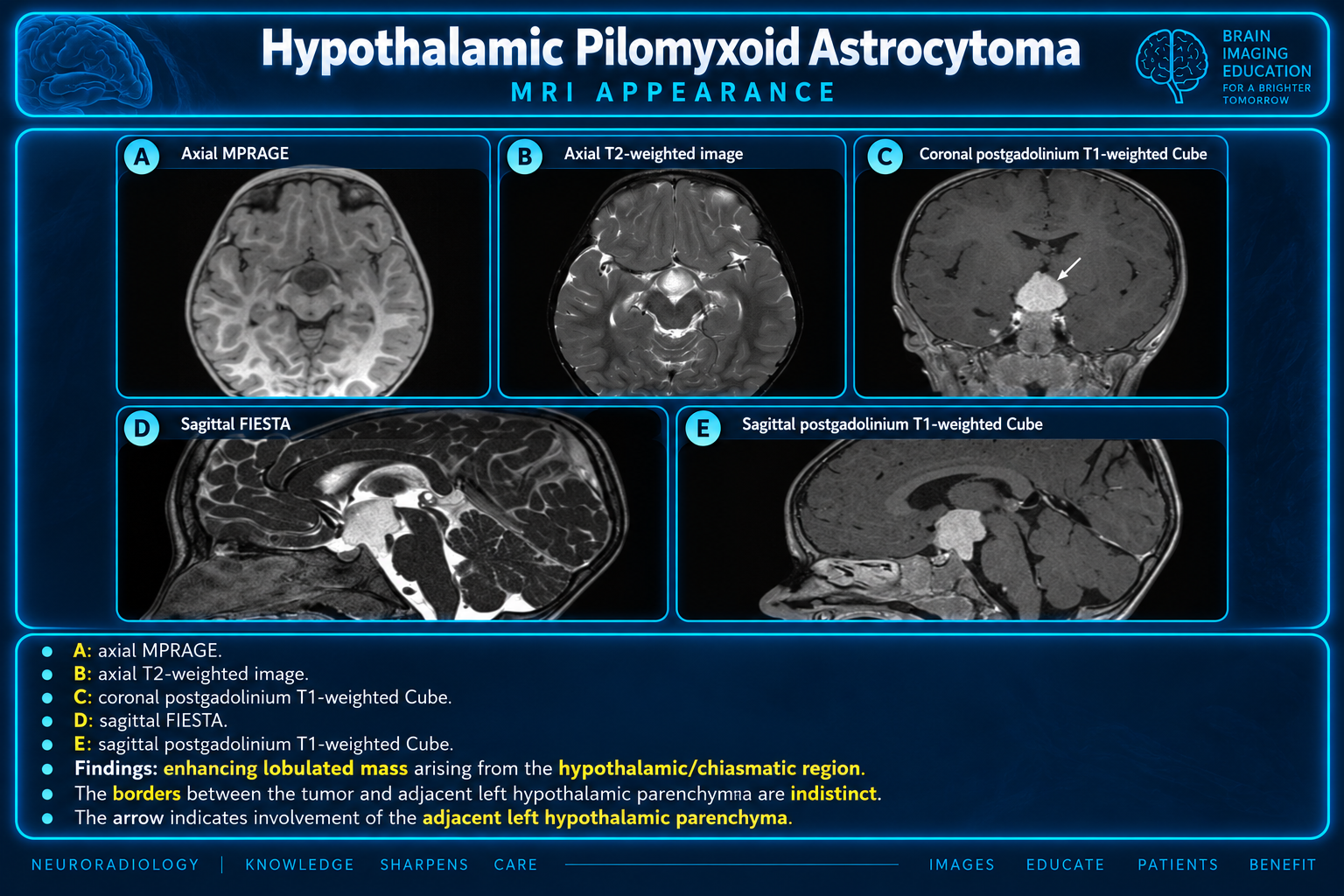
Pilomyxoid astrocytoma is a histologic pattern within the pilocytic astrocytoma spectrum. It classically affects infants and young children in the hypothalamic-chiasmatic region.
- Predominantly solid, well-circumscribed mass.
- Very homogeneous T2 hyperintensity and relatively homogeneous enhancement.
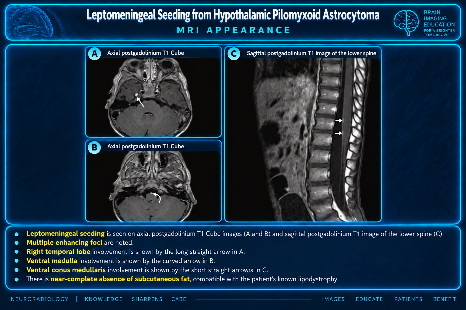
- Greater tendency toward hemorrhage, hydrocephalus, recurrence, and CSF dissemination.
- Imaging cannot reliably establish the histologic pattern.
Dissemination
Leptomeningeal nodules or ependymal disease should prompt evaluation of the entire neuraxis. Spine MRI is particularly important when dissemination is suspected or a pilomyxoid pattern is established.
- Inspect basal cisterns and ventricular surfaces.
- Look for enhancing intracranial and spinal nodules.
- Compare for new hydrocephalus or ependymal disease.
CT and Calcification
CT may show a low- or isoattenuating suprasellar-hypothalamic mass and is useful for hemorrhage, hydrocephalus, calcification, and osseous anatomy.
Diagnostic redirectProminent calcification is much more characteristic of craniopharyngioma than optic pathway-hypothalamic glioma and should prompt reconsideration of the diagnosis.
Key Differential Diagnosis
| Entity | Distinguishing imaging clues |
|---|
| Germinoma | Vertical stalk-centered mass, relatively T2 low signal, avid homogeneous enhancement, restricted diffusion, absent posterior bright spot, and possible synchronous pineal lesion |
| Craniopharyngioma | Mixed solid-cystic mass; calcification favors the adamantinomatous subtype; cyst contents may be intrinsically T1 hyperintense |
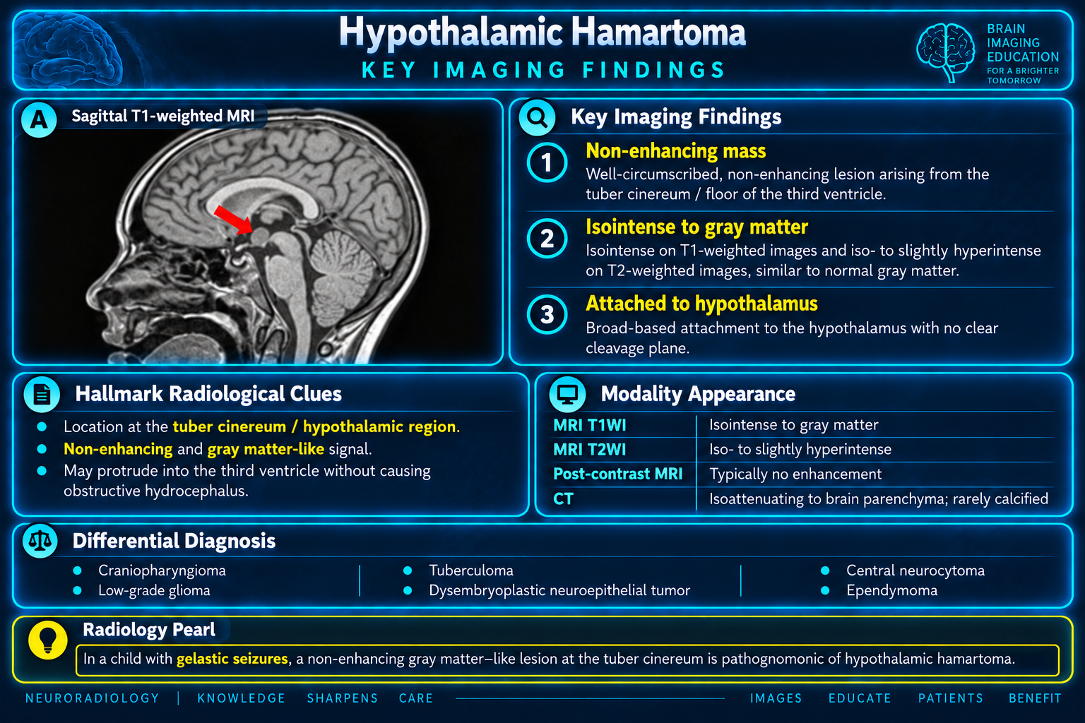
| Hypothalamic hamartoma | Stable tuber cinereum lesion that follows gray matter, does not enhance or restrict, and does not expand the optic pathway |
| LCH, hypophysitis, or sarcoidosis | Predominantly stalk-centered thickening, absent posterior bright spot, diabetes insipidus, and systemic or meningeal findings |
| Lymphoma | Relatively T2-dark, diffusion-restricting, avidly enhancing lesion with possible periventricular or meningeal disease |
| Chordoid glioma | Adult anterior third-ventricular mass, usually avidly enhancing without optic pathway expansion |
| Metastasis | Older patient, known malignancy, rapid growth, prominent enhancement or edema, and other metastases |
| High-grade optic pathway glioma | Adult with rapid visual decline, infiltrative chiasm-tract expansion, necrosis, edema, and heterogeneous or ring enhancement |
Stalk Disease Distinction
Establish whether the geometric center is the chiasm-hypothalamus or the infundibulum.
- A T2-bright expansile mass continuous with the chiasm or optic tracts favors glioma.
- Isolated smooth infundibular thickening without optic pathway expansion is atypical.
- The stalk may be displaced, engulfed, or directly invaded by a large glioma.
MRI Protocol
- Thin 3D pre- and postcontrast T1 with multiplanar reformations.
- Axial and coronal T2/FLAIR through the hypothalamic-optic pathways.
- Small-field-of-view fat-suppressed orbital T2 and postcontrast T1 when optic nerve disease is possible.
- DWI/ADC and SWI/GRE.
- Perfusion or spectroscopy for atypical or progressive lesions.
- Whole-brain and spine postcontrast imaging when dissemination is suspected.
Reporting Checklist
- Each optic nerve segment, chiasm, tracts, lateral geniculate bodies, and optic radiations.
- Hypothalamic side, tuber cinereum, mammillary region, and third-ventricular floor.
- Relationship to the pituitary stalk.
- Suprasellar, interpeduncular, prepontine, and temporal extension.
- Vascular encasement or displacement.
- Hydrocephalus, hemorrhage, calcification, restriction, enhancement, and dissemination.
Follow-up Interpretation
- Measure enhancing and nonenhancing disease on consistent sequences and planes.
- T2/FLAIR extent is often more reliable than enhancement.
- Do not call progression from increased enhancement alone without corroborating growth.
- Assess cyst enlargement, hemorrhage, hydrocephalus, new restriction, necrosis, or dissemination.
- Correlate with visual fields, endocrine status, growth, and hypothalamic symptoms.
Practical Impression Language
Typical low-grade pattern
Lobulated expansile T2/FLAIR-hyperintense mass centered in the optic chiasm and hypothalamus, with extension along the [right/left/bilateral] optic tract or nerve and [enhancement pattern]. Facilitated diffusion and absence of substantial edema or necrosis favor a low-grade optic pathway-hypothalamic glioma, most commonly pilocytic astrocytoma in this age group. Correlate for NF1 and visual or endocrine dysfunction.
Pilomyxoid-pattern concern
Predominantly solid, avidly enhancing hypothalamic-chiasmatic mass with homogeneous T2 hyperintensity and associated [finding]. The young age, solid morphology, and [leptomeningeal dissemination/hemorrhage/hydrocephalus] raise consideration of a pilomyxoid pattern, although imaging cannot reliably distinguish it from conventional pilocytic astrocytoma.
Atypical adult pattern
Infiltrative mass expanding the optic chiasm, tracts, and hypothalamus with heterogeneous enhancement, necrosis, and [finding]. In an adult with rapidly progressive visual symptoms, aggressive optic pathway glioma is a consideration; germinoma, lymphoma, metastasis, and inflammatory or infiltrative disease remain important alternatives. Tissue diagnosis and multidisciplinary evaluation are recommended.
Key Takeaways
- Most pediatric lesions belong to the optic pathway-hypothalamic glioma spectrum.
- The signature combination is a T2/FLAIR-bright expansile chiasmatic-hypothalamic mass with facilitated diffusion.
- Longitudinal optic pathway involvement matters more than enhancement intensity.
- Solid homogeneous tumors with hemorrhage, hydrocephalus, or dissemination raise a pilomyxoid pattern.
- Marked restriction, calcification, or a nonenhancing gray-matter-like lesion should redirect the differential.
Related Pages