External Auditory Canal Masses

Imaging approach: Begin with high-resolution temporal-bone CT to determine whether the canal finding is debris, bone, a circumscribed soft-tissue lesion, or an infiltrative mass. The pattern of bone response—none, smooth remodeling, focal erosion, or aggressive destruction—is the most useful initial discriminator. Add contrast-enhanced MRI when tumor, skull-base infection, marrow invasion, dural extension, or perineural spread is suspected.

Visual Reference

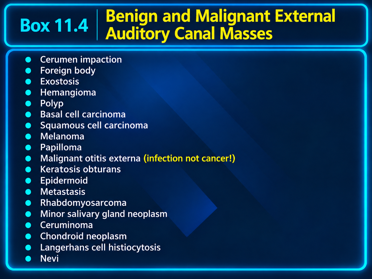
Benign and malignant external auditory canal mass differential
External Auditory Canal Mass DifferentialClick to enlarge

Obstructing Mimics

Usually no bone erosionCorrelate with otoscopy before interpreting canal debris as a true mass.

Cerumen and foreign body

Cerumen is nonenhancing, often nonadherent debris with peripheral air or internal gas. Foreign material may be directly visible when radiopaque; chronic cases can develop edema or granulation tissue.

Keratosis obturans

Keratin fills and smoothly widens the bony canal, often bilaterally, without focal erosion or osseous sequestra. The middle ear and mastoid are usually preserved.

Aural polyp

A nonspecific inflammatory mass associated with chronic infection, cholesteatoma, foreign-body reaction, or surgery. Bone erosion should prompt a search for the underlying cause rather than a simple “polyp” diagnosis.

Benign Lesions

Circumscribed or smoothly remodelingAggressive irregular destruction is not a typical benign response.
  • Exostoses: multiple broad-based medial bony protuberances, commonly bilateral and related to cold-water exposure.
  • Osteoma: typically solitary, unilateral, and pedunculated.
  • Hemangioma/venous malformation: avidly enhancing vascular mass; may contain phleboliths.
  • Papilloma: superficial circumscribed nodule without osseous erosion.
  • Epidermoid cyst: well-defined keratin-containing mass with diffusion restriction.
  • Nevus: small superficial skin lesion; growth, ulceration, bleeding, or deep invasion requires exclusion of melanoma.

Infection and Malignancy

Aggressive EAC processIrregular bone destruction and deep extension raise concern for carcinoma or necrotizing otitis externa.

Necrotizing otitis externa

Invasive infection, not cancer. Look for canal-wall inflammation, tympanic-bone erosion, skull-base osteomyelitis, deep facial-space spread, and cranial neuropathy—especially in an older patient with diabetes or immunocompromise.

Squamous cell carcinoma

The dominant primary EAC malignancy. Persistent otorrhea, pain, bleeding, a nonhealing polyp, facial weakness, or trismus should prompt evaluation for an infiltrative mass with irregular bone destruction.

Other malignancies

BCC, melanoma, metastasis, pediatric rhabdomyosarcoma, ceruminous or other salivary-type tumors, chondroid neoplasm, and Langerhans cell histiocytosis.
Bone-Response Pattern

PatternLeading considerations
No erosionCerumen, foreign body, uncomplicated polyp, most benign superficial lesions
Smooth generalized wideningKeratosis obturans; chronic pressure from a slow-growing benign lesion
Multiple smooth bony projectionsExostoses; solitary pedunculated lesion favors osteoma
Focal erosion with keratin/debrisExternal auditory canal cholesteatoma
Irregular destructive erosionSCC, necrotizing otitis externa, other carcinoma, metastasis, sarcoma, LCH

CT and MRI Checklist

High-resolution CT

  • Cartilaginous versus bony canal origin.
  • Anterior, posterior, superior, or inferior canal wall involvement.
  • Smooth remodeling versus focal or irregular erosion.
  • Tympanic membrane, middle ear, ossicles, mastoid, TMJ, and facial canal.

Contrast-enhanced MRI

  • Enhancement, diffusion restriction, and marrow invasion.
  • Parotid, masticator-space, skull-base, dural, and intracranial extension.
  • Facial nerve or other perineural involvement.

Important Specific Diagnoses

  • EAC cholesteatoma: focal canal-wall erosion with keratin debris; distinguish from the smooth widening of keratosis obturans.
  • Melanoma: may be T1 hyperintense and T2 hypointense when melanotic, but imaging appearance is variable.
  • Ceruminous tumors: arise in the outer cartilaginous canal; replace the nonspecific term “ceruminoma” with a histologic diagnosis when available.
  • Chondroid tumors: ring-and-arc or stippled matrix on CT; cartilage-rich tissue is often T2 bright.
  • LCH: sharply marginated lytic temporal-bone lesion with canal or scalp soft tissue, often in children.
  • Metastasis: consider with known systemic malignancy, multifocal bone lesions, or a hypervascular mass.

Reporting Language

Exostoses: “Broad-based bilateral smooth osseous protuberances of the medial bony external auditory canals, producing narrowing without erosion or soft-tissue mass.”
Keratosis obturans: “Smooth generalized EAC expansion by nonenhancing keratinous debris, without focal osseous erosion or middle-ear disease.”
Suspected malignancy: “Enhancing infiltrative EAC mass with irregular tympanic-bone erosion and extension toward the TMJ/parotid space. MRI is recommended for skull-base, marrow, dural, and perineural staging.”
Suspected invasive infection: “EAC inflammatory soft tissue with temporal-bone/skull-base marrow abnormality and deep-space inflammatory enhancement; necrotizing otitis externa is favored in the appropriate clinical setting.”

Secondary Links