Imaging clue
Longitudinal, sometimes tract-selective T2 signal, particularly in dorsal columns or lateral tracts; this is not specific.
An imaging reference for spinal cord involvement, differential diagnosis, and reporting. There is no diagnostic MRI signature; attribution requires clinical and laboratory correlation.
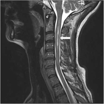
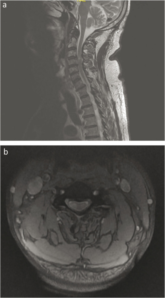
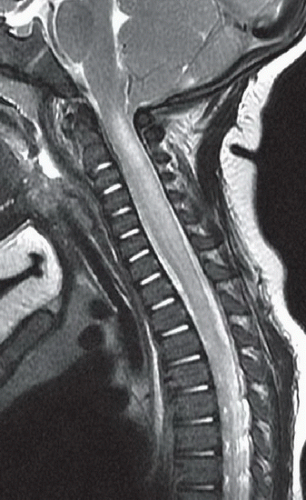
Source limitations: the second graphic has a discordant “ARIA-E” header. Images 3–4 have no supplied diagnostic captions or patient context; they are not independently established examples of Sjögren myelitis. Image 5 retains the supplied caption.
Longitudinal, sometimes tract-selective T2 signal, particularly in dorsal columns or lateral tracts; this is not specific.
Sjögren disease can coexist with AQP4-positive NMOSD. Sjögren status alone does not establish the cause of a cord lesion.
A normal early MRI or absent enhancement does not exclude inflammatory disease. Posterior-column signal may also reflect sensory neuronopathy.
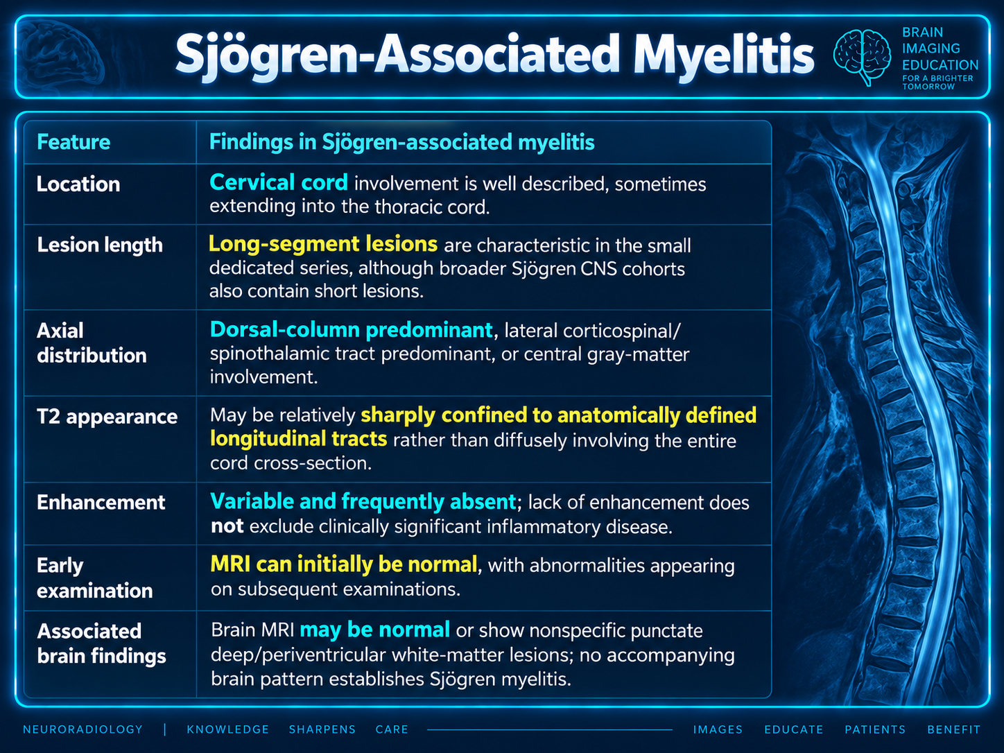
Focusing just on Sjögren-associated myelitis: it is a rare inflammatory spinal cord manifestation that can occur independently of NMOSD. The most useful radiologic clue is longitudinal, sometimes tract-selective T2 hyperintensity—particularly involving the dorsal columns or lateral tracts—but there is no diagnostic MRI signature. pmc.ncbi.nlm.nih
| Feature | Findings in Sjögren-associated myelitis |
|---|---|
| Location | Cervical cord involvement is well described, sometimes extending into the thoracic cord. pmc.ncbi.nlm.nih |
| Lesion length | Long-segment lesions are characteristic in the small dedicated series, although broader Sjögren CNS cohorts also contain short lesions. pmc.ncbi.nlm.nih |
| Axial distribution | Dorsal-column predominant, lateral corticospinal/spinothalamic tract predominant, or central gray-matter involvement. pmc.ncbi.nlm.nih |
| T2 appearance | May be relatively sharply confined to anatomically defined longitudinal tracts rather than diffusely involving the entire cord cross-section. pmc.ncbi.nlm.nih |
| Enhancement | Variable and frequently absent; lack of enhancement does not exclude clinically significant inflammatory disease. pmc.ncbi.nlm.nih |
| Early examination | MRI can initially be normal, with abnormalities appearing on subsequent examinations. pmc.ncbi.nlm.nih |
| Associated brain findings | Brain MRI may be normal or show nonspecific punctate deep/periventricular white-matter lesions; no accompanying brain pattern establishes Sjögren myelitis. pmc.ncbi.nlm.nih |
A particularly relevant study is Butryn et al., Journal of Clinical Medicine, 2020, which described four patients with Sjögren-associated inflammatory myelitis: all were AQP4- and MOG-antibody negative, and none met NMOSD criteria. pmc.ncbi.nlm.nih
The caveat: this is a four-patient series, so tract-selective involvement is a useful consideration—not a validated discriminator or a finding specific to Sjögren disease. pmc.ncbi.nlm.nih
For a compatible lesion in someone with established Sjögren disease, reasonable wording would be:
“Longitudinally extensive, [dorsal-column/lateral-tract/central]-predominant intramedullary T2 signal abnormality, compatible with inflammatory myelitis. Sjögren-associated myelitis is a consideration given the clinical history; the imaging appearance is not specific.”
Sjögren-associated myelitis has no single diagnostic MRI pattern. In a patient with Sjögren disease and myelitis—particularly longitudinally extensive transverse myelitis (LETM)—the primary radiologic concern should be coexisting AQP4-IgG–positive neuromyelitis optica spectrum disorder (NMOSD) rather than automatically attributing the lesion to Sjögren disease itself. pmc.ncbi.nlm.nih
The report covers:
The most NMOSD-favoring findings are central LETM, cord expansion, bright spotty lesions, cervicomedullary extension, posterior/chiasmal optic neuritis, and area-postrema or AQP4-rich periependymal brain lesions. Serum AQP4-IgG testing by cell-based assay—and MOG-IgG testing if negative—is essential because Sjögren disease can coexist with NMOSD, while AQP4/MOG-negative Sjögren myelitis remains a diagnosis of exclusion. pmc.ncbi.nlm.nih
Even when focusing on Sjögren itself, the diagnosis should remain clinicoradiologic rather than imaging-only: Sjögren status does not prove the cause of a cord lesion, and AQP4/MOG-associated disease should still be excluded before assigning it directly to Sjögren disease. pmc.ncbi.nlm.nih
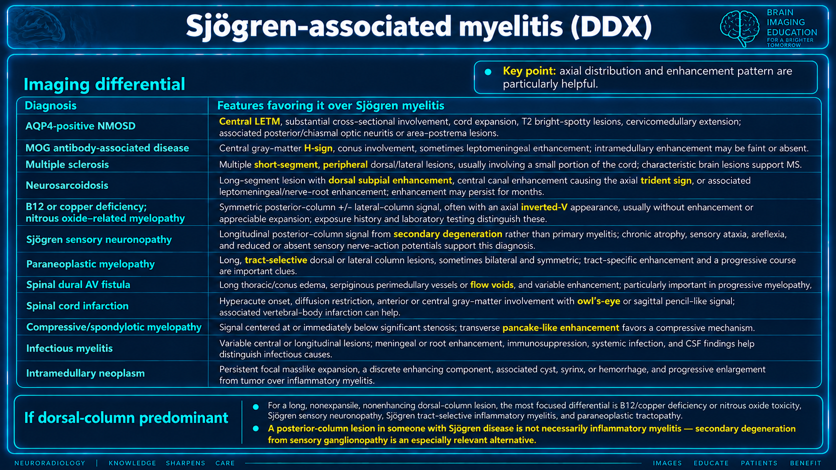
For suspected Sjögren-associated myelitis, the main radiologic differential includes inflammatory demyelination, metabolic or degenerative tractopathy, and vascular mimics; axial distribution and enhancement pattern are particularly helpful. pmc.ncbi.nlm.nih
| Diagnosis | Features favoring it over Sjögren myelitis |
|---|---|
| AQP4-positive NMOSD | Central LETM, substantial cross-sectional involvement, cord expansion, T2 “bright spotty” lesions, cervicomedullary extension; associated posterior/chiasmal optic neuritis or area-postrema lesions. pmc.ncbi.nlm.nih |
| MOG antibody-associated disease | Central gray-matter H-sign, conus involvement, and sometimes leptomeningeal enhancement; intramedullary enhancement may be faint or absent. pmc.ncbi.nlm.nih |
| Multiple sclerosis | Multiple short-segment, peripheral dorsal/lateral lesions, usually involving a small portion of the cord; accompanying characteristic brain lesions support MS. pmc.ncbi.nlm.nih |
| Neurosarcoidosis | Long-segment lesion with dorsal subpial enhancement, central canal enhancement producing the axial trident sign, or associated leptomeningeal/nerve-root enhancement; enhancement may persist for months. pmc.ncbi.nlm.nih |
| B12 or copper deficiency; nitrous oxide–related myelopathy | Symmetric posterior-column ± lateral-column signal, often an axial inverted-V appearance, usually without enhancement or appreciable expansion; exposure history and laboratory testing distinguish these. pmc.ncbi.nlm.nih |
| Sjögren sensory neuronopathy | Longitudinal posterior-column signal from secondary degeneration rather than primary myelitis; chronic atrophy, sensory ataxia, areflexia, and reduced/absent sensory nerve-action potentials support this diagnosis. pmc.ncbi.nlm.nih |
| Paraneoplastic myelopathy | Long, tract-selective dorsal or lateral column lesions, sometimes bilateral and symmetric; tract-specific enhancement and a progressive course are important clues. pmc.ncbi.nlm.nih |
| Spinal dural AV fistula | Long thoracic/conus edema, serpiginous perimedullary vessels/flow voids, and variable enhancement; particularly important in a progressive myelopathy. pmc.ncbi.nlm.nih |
| Spinal cord infarction | Hyperacute onset, diffusion restriction, anterior/central gray-matter involvement with owl’s-eye or sagittal pencil-like signal; associated vertebral-body infarction can help. pmc.ncbi.nlm.nih |
| Compressive/spondylotic myelopathy | Signal centered at or immediately below significant stenosis; transverse pancake-like enhancement favors a compressive mechanism. pmc.ncbi.nlm.nih |
| Infectious myelitis | Variable central or longitudinal lesions; meningeal/root enhancement, immunosuppression, systemic infection, and CSF findings help distinguish viral, syphilitic, tuberculous, or other infectious causes. pmc.ncbi.nlm.nih |
| Intramedullary neoplasm | Persistent focal masslike expansion, a discrete enhancing component, associated cyst/syrinx or hemorrhage, and progressive enlargement favor tumor over inflammatory myelitis. pmc.ncbi.nlm.nih |
For a long, nonexpansile, nonenhancing dorsal-column lesion, the most focused differential is B12/copper deficiency or nitrous oxide toxicity, Sjögren sensory neuronopathy, Sjögren tract-selective inflammatory myelitis, and paraneoplastic tractopathy. pmc.ncbi.nlm.nih
A posterior-column lesion in someone with Sjögren disease is not necessarily inflammatory myelitis—secondary degeneration from sensory ganglionopathy is an especially relevant alternative. jnnp.bmj
Scope limitation: the supplied text announces a recommended MRI protocol and broader brain/orbital review, but does not provide a complete protocol or detailed review. No additional protocol has been inferred.