US-Guided FNA for Salivary Gland Masses
Reference Images
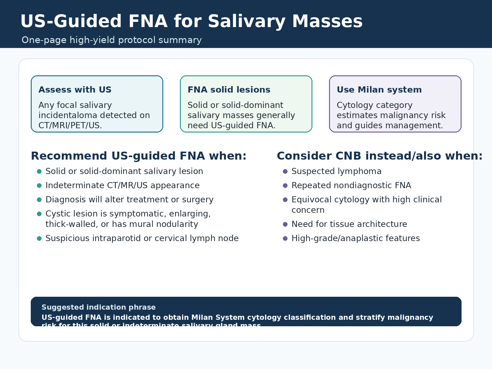
FNA Indications
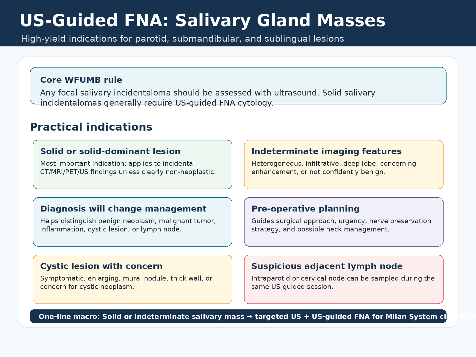
Core rule
Any focal salivary incidentaloma should be assessed with ultrasound. Solid or solid-dominant salivary incidentalomas generally require US-guided FNA cytology.
Recommend US-guided FNA when
- Solid or solid-dominant salivary lesion.
- Indeterminate CT, MRI, PET, or ultrasound appearance.
- Diagnosis will alter treatment, surgery, or follow-up.
- Cystic lesion is symptomatic, enlarging, thick-walled, has mural nodularity, or has a solid component.
- Suspicious intraparotid or cervical lymph node can be sampled during the same ultrasound-guided session.
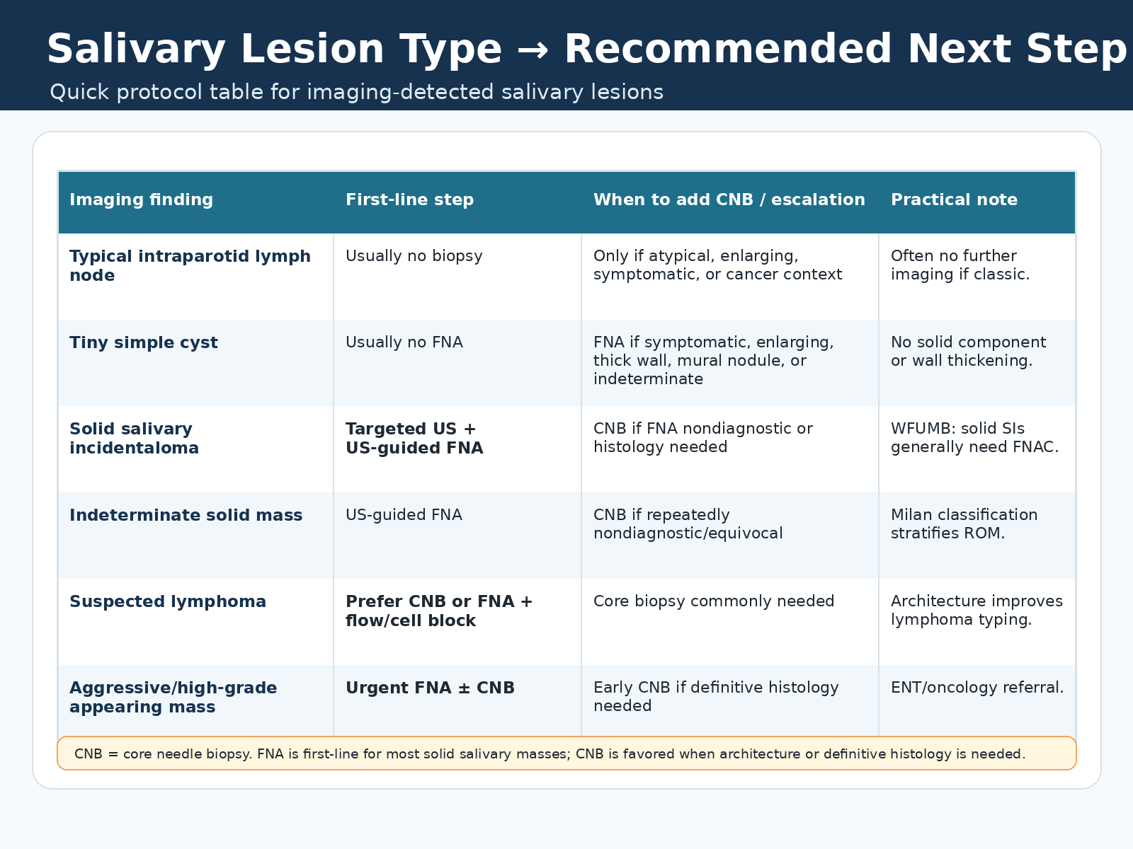
Macro: Solid or indeterminate salivary mass - targeted ultrasound plus US-guided FNA for Milan System cytology classification.
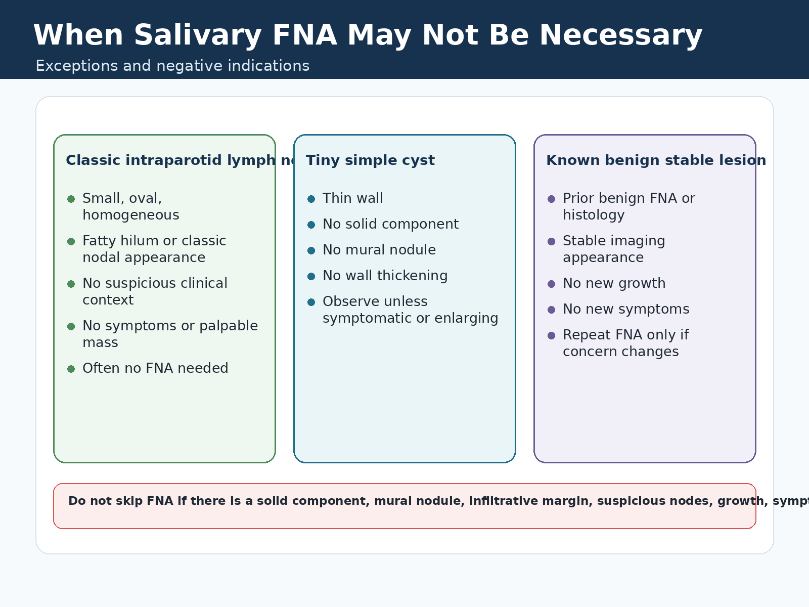
When FNA May Not Be Needed
Often observe / no biopsy
- Classic intraparotid lymph node: small, oval, homogeneous, fatty hilum or classic nodal appearance, and no suspicious clinical context.
- Tiny simple cyst: thin wall, no solid component, no mural nodule, and no wall thickening.
- Known benign stable lesion: prior benign FNA or histology, stable imaging appearance, no growth, and no new symptoms.
- Repeat FNA is usually reserved for interval growth or a change in clinical/imaging concern.
Do not skip FNA if
- Solid component, mural nodule, infiltrative margin, suspicious nodes, growth, symptoms, or malignancy context.
- Cystic lesion is symptomatic, enlarging, thick-walled, or concerning for cystic neoplasm.
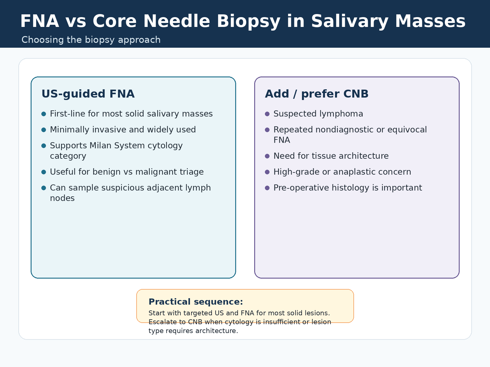
FNA vs CNB
FNA first-line
- First-line for most solid salivary masses.
- Minimally invasive and widely used for benign versus malignant triage.
- Supports Milan System cytology category and risk stratification.
- Can sample suspicious adjacent intraparotid or cervical lymph nodes.
Add or prefer CNB when
- Suspected lymphoma or need for tissue architecture.
- Repeated nondiagnostic or equivocal FNA.
- High-grade or anaplastic concern.
- Definitive pre-operative histology is important for management.