Age-related white matter hyperintensities (WMH) are common MRI findings that usually reflect cerebral small vessel disease (SVD), particularly arteriolosclerotic disease of deep perforating vessels. They are not a diagnosis by themselves: relevance depends on age, vascular risk profile, lesion burden and distribution, symptoms, and accompanying SVD markers.
WMH are one manifestation in a spectrum that includes recent small subcortical infarcts, lacunes, enlarged perivascular spaces, cerebral microbleeds, cortical superficial siderosis, microinfarcts, and brain atrophy. Small punctate foci are common in older adults; confluent, progressive, or disproportionate disease is more meaningful.
Terminology
Cerebral small vessel disease is widely defined in the literature.
With respect to anatomy, small vessels include small arteries, arterioles, capillaries, venules, and small veins. The arterial bed has been a primary focus of the literature, with arterial small vessel disease being a proposed term for this entity, but venous collagenosis is also responsible for some of the changes 15. The small vessels involved are too small to visualize by imaging in vivo, so imaging focuses on their sequelae.
With respect to morphologic changes visible on imaging, cerebral small vessel disease includes 14,16,20:
Although white matter hyperintensities are a key imaging marker of small vessel disease, they are not specific to it, and other causes of white matter signal abnormality should be considered in clinical context. Analogous signal changes in the subcortical grey matter and brainstem are typically excluded from discussion unless explicitly stated, leading to terms that focus on small vessel disease of white matter 16,20.
The term leukoaraiosis is a radiological descriptor applied to white matter hypodensities on CT and high signal changes on T2-weighted MRI of presumed vascular origin 14,16. These lesions are properly termed white matter hyperintensities (WMH) on MRI, as defined by Standards for Reporting Vascular Changes on Neuroimaging 2 (STRIVE-2), but are also commonly referred to as white matter lesions or white matter changes 16,20. Similar terms focused on the white matter include white matter disease, white matter damage, and leukoencephalopathy (the latter usually used in the context of CADASIL) 16,20. Less commonly, the lesions are called unidentified bright objects on MRI, but this term has also confusingly been used to refer to the focal areas of signal intensity in brains of children with neurofibromatosis type 1, which is an unrelated process.
Many etiopathogenic types of small vessel disease are described. The most common is arteriolosclerosis, or age- and vascular-risk-factor-related small vessel disease, which—based on a progressive clinical syndrome of cognitive impairment and compatible imaging features—is diagnosed as Binswanger disease 14, although this term has fallen in popularity. Many variant terms presume an age-related etiology, such as age-related white matter hyperintensities, changes, disease, or damage 16, although not all age-related white matter changes are attributed to small vessel disease 14.
The nature of infarcts and white matter changes is primarily ischemic, so other terms include small vessel chronic ischemia, microvascular ischemia, ischemic microangiopathy, and variants such as ischemic white matter disease. However, hemorrhagic manifestations—including cerebral microbleeds, intracerebral hemorrhage, and cortical superficial siderosis—are also important for differential diagnosis and therapeutic decisions.
Terminology and Scope
| Term | Preferred meaning / practical use |
|---|
| Cerebral small vessel disease (SVD) | Umbrella term for brain injury attributable to small arteries, arterioles, capillaries, venules, or small veins; imaging depicts downstream injury. |
| White matter hyperintensities (WMH) | T2/FLAIR hyperintense white matter lesions, ideally qualified as “of presumed vascular origin” when appropriate. |
| Leukoaraiosis | Historical term for CT hypodensity or T2/FLAIR hyperintensity, usually presumed vascular. |
| Chronic microvascular ischemic change | Common reporting phrase for a presumed vascular pattern; WMH remain pathologically nonspecific. |
| Deep perforator arteriopathy | Age- and risk-factor-related arteriolar disease associated with deep WMH, lacunes, and deep microbleeds. |
| Cerebral amyloid angiopathy (CAA) | Cortical/leptomeningeal vasculopathy associated with lobar microbleeds, superficial siderosis, and convexity SAH. |
| Leukoencephalopathy | Broad term often reserved for inherited, inflammatory, toxic, metabolic, or other nonroutine patterns. |
“White matter disease” and “white matter changes” are descriptive, not etiologic. FLAIR hyperintensity alone does not prove chronic ischemia.
Pathobiology
Histologic substrate
- Myelin pallor/loss and axonal loss
- Oligodendrocyte injury
- Increased interstitial water
- Astrocytosis and microglial activation
- Enlarged perivascular spaces
- Small infarcts or microinfarcts
Vascular mechanism
- Arteriolosclerosis and lipohyalinosis
- Wall thickening and luminal narrowing
- Impaired autoregulation, hypoperfusion, and oxygen delivery
Beyond ischemia
- Endothelial dysfunction and BBB leakage
- Inflammation, altered fluid handling, venous collagenosis
- Oxidative stress and declining myelin repair
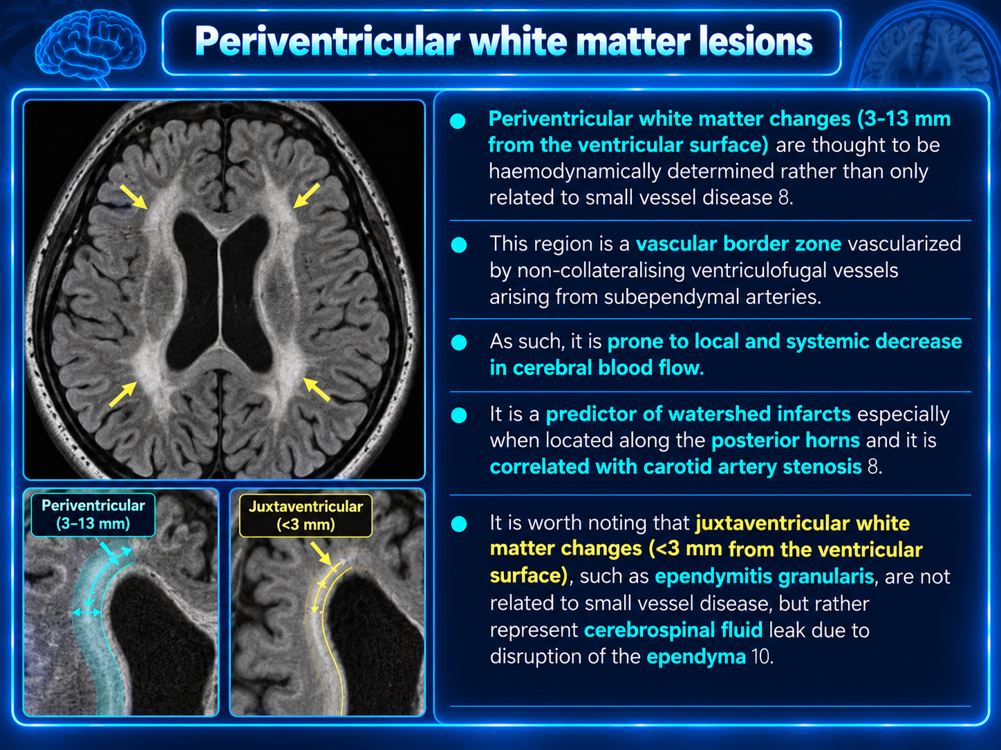
A thin smooth periventricular cap may partly reflect ependymal disruption and interstitial fluid rather than consequential ischemia.
Imaging Assessment
Preferred MRI protocol: T1, T2, FLAIR, DWI/ADC, and GRE or SWI; add vascular imaging when indicated.
- FLAIR/T2: hyperintense.
- T1: iso- to mildly hypointense; more hypointense with severe injury.
- DWI/ADC: no restriction when chronic.
- Postcontrast: usually no enhancement.
- CT: ill-defined or confluent hypoattenuation when advanced.
Distribution
- Juxtaventricular caps may reflect ependymal/interstitial change.
- Periventricular disease may reflect border-zone vulnerability.
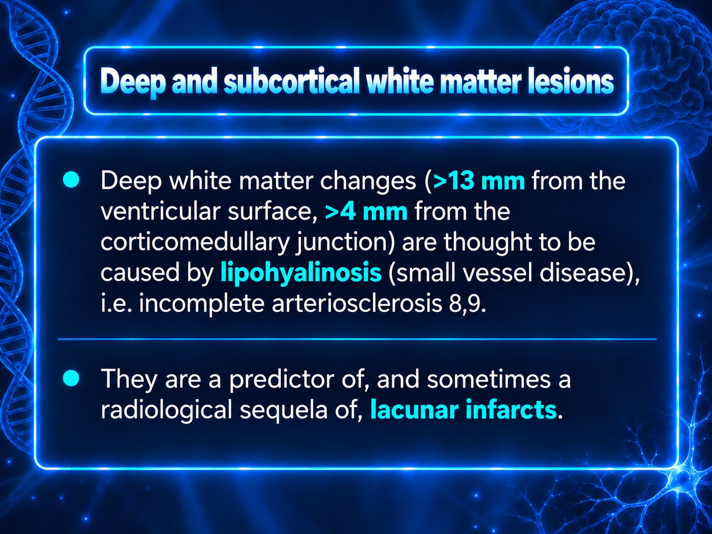
- Deep WMH favor arteriolosclerotic perforator disease.
- Marked U-fiber disease is atypical.
- Asymmetric, mass-like, enhancing, or restricting lesions require broader differential.
Associated SVD Markers
- Recent small subcortical infarct
- Lacune of presumed vascular origin
- Enlarged perivascular spaces
- Cerebral microbleeds
- Cortical superficial siderosis
- Convexity subarachnoid hemorrhage
- Cortical microinfarcts
- Disproportionate or progressive atrophy
Deep/infratentorial microbleeds favor hypertensive deep perforator arteriopathy. Strictly lobar microbleeds and superficial siderosis raise concern for CAA.
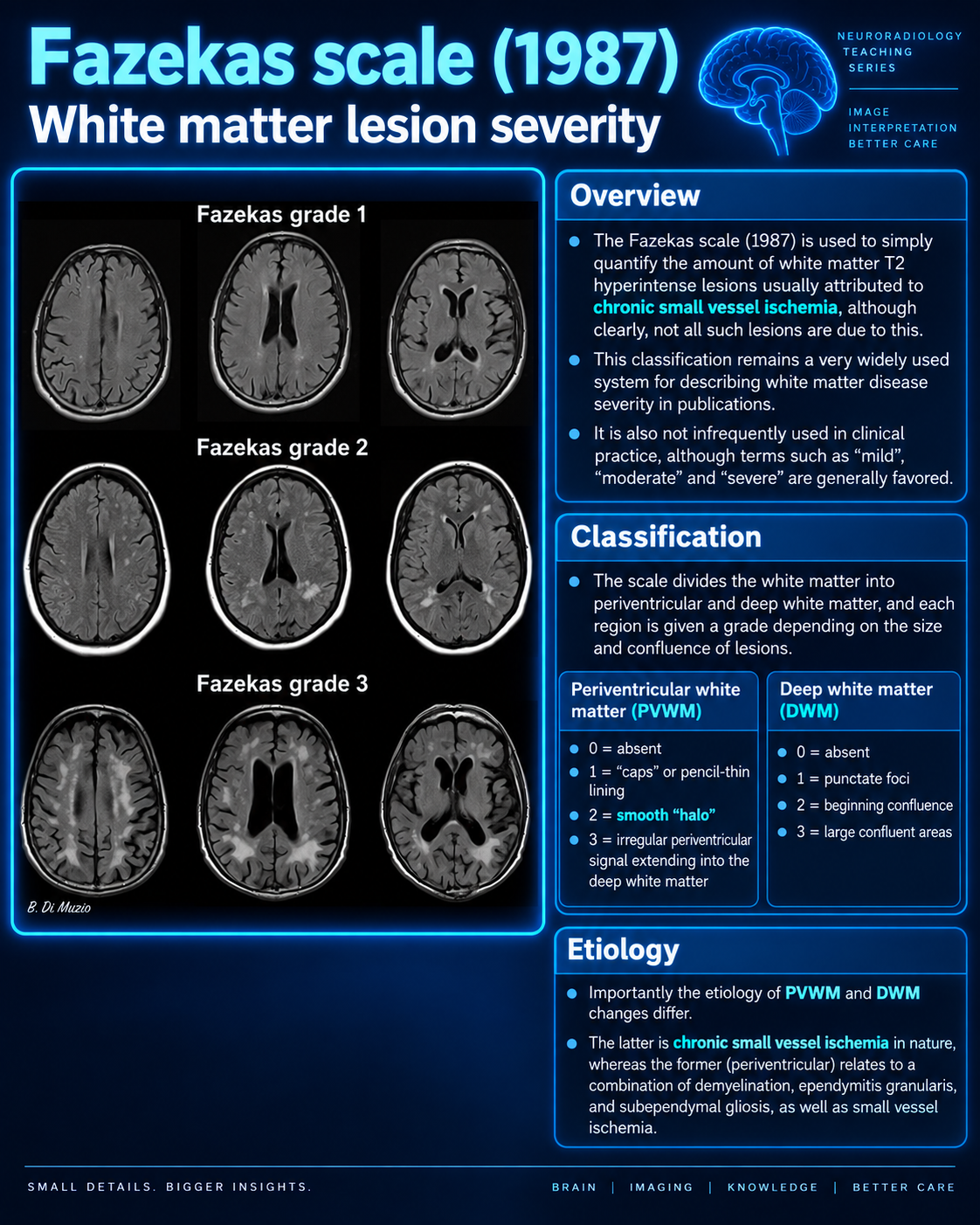
Fazekas Visual Scale
| Grade | Periventricular WMH | Deep WMH | Practical interpretation |
|---|
| 0 | None | None | No visible WMH |
| 1 | Caps or thin lining | Punctate foci | Common with aging; often limited significance in isolation |
| 2 | Smooth halo | Beginning confluence | Moderate burden; assess risk factors and SVD markers |
| 3 | Irregular extension into deep white matter | Large confluent areas | Advanced burden; greater cognitive, gait, and cerebrovascular risk |
Score periventricular and deep WMH separately. Volumetry is more sensitive for research and longitudinal change, but Fazekas remains practical clinically.
Clinical Importance
Associations are probabilistic and modified by location, reserve, neurodegeneration, infarcts, microbleeds, education, and activity.
- Slowed processing and executive dysfunction
- MCI and increased dementia risk
- Gait slowing, instability, and falls
- Urinary urgency/incontinence in advanced disease
- Depression and apathy
- Increased ischemic stroke risk
- Hemorrhagic risk in appropriate phenotypes
WMH can coexist and interact with Alzheimer disease but are not synonymous with it.
Risk Factors
Age is the strongest correlate; hypertension is the most important modifiable driver.
- Hypertension, especially cumulative midlife exposure
- Diabetes/insulin resistance
- Smoking, obesity, and inactivity
- Sleep disturbance or suspected sleep apnea
- Excess alcohol and poor diet
- Depression, isolation, and low engagement
LDL alone is not an established direct cause, although lipid management matters for overall cardiovascular risk. Genetics, endothelial biology, inflammation, resilience, and neurodegeneration also contribute.
Red Flags / Differential
- Young with extensive WMH: genetic SVD, CADASIL, inflammatory, migraine, toxic-metabolic, treatment-related disease.
- Anterior temporal/external capsule: CADASIL pattern.
- Asymmetric or tumefactive: neoplasm, demyelination, infection, subacute infarct.
- Callosal, juxtacortical, infratentorial, or cord-predominant: inflammatory/demyelinating disease.
- Posterior vasogenic edema: consider PRES.
- Lobar microbleeds/siderosis/convexity SAH: CAA.
- Enhancement or restriction: active process, not routine chronic WMH.
- Radiation, chemotherapy, toxin, immunosuppression: treatment-related, toxic, infectious, or metabolic disease.
Management Framework
No established drug reverses typical age-related WMH. Reduce further injury and address meaningful symptoms.
- Individualize and optimize blood pressure.
- Address diabetes, obesity, inactivity, and metabolic health.
- Stop smoking and avoid excess alcohol.
- Encourage aerobic and resistance exercise.
- Use Mediterranean- or DASH-style eating patterns.
- Evaluate focal deficits, stepwise decline, gait, cognitive, or urinary symptoms.
- Base antiplatelet/statin use on standard indications—not incidental WMH alone.
- Consider hemorrhagic markers in antithrombotic decisions.
- Use formal cognitive testing when symptoms affect function, work, or driving.
Practical Reporting Language
Mild burden
Mild scattered supratentorial white matter T2/FLAIR hyperintensities, nonspecific but most commonly related to chronic microvascular ischemic change.
Moderate burden
Moderate patchy and confluent supratentorial white matter T2/FLAIR hyperintensity, most consistent with chronic small vessel ischemic change. Consider correlation with vascular risk factors.
Advanced burden
Extensive confluent periventricular and deep cerebral white matter T2/FLAIR hyperintensity, compatible with advanced chronic small vessel ischemic disease. Associated chronic lacunar infarcts and/or microhemorrhages, if present, should be separately described.
When caution is appropriate
White matter signal abnormality is greater than expected for age and/or demonstrates an atypical distribution. Correlate with clinical history and consider alternative inflammatory, demyelinating, hereditary, toxic-metabolic, or other leukoencephalopathic etiologies as appropriate.