Supraglottic Cancer

Core concept: Supraglottic squamous cell carcinoma may have a modest mucosal footprint but substantial submucosal extension. Imaging complements endoscopy by mapping the pre-epiglottic and paraglottic spaces, cartilage, tissues beyond the larynx, airway, and bilateral cervical nodal chains.

Visual Reference

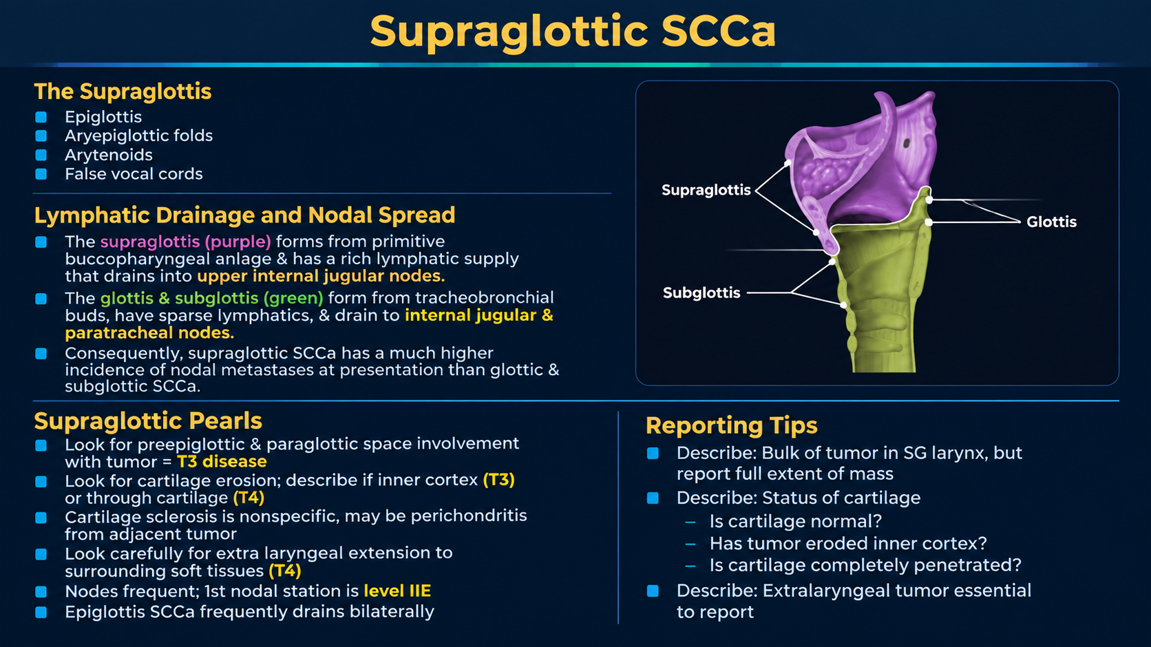
Supraglottic SCC overview and nodal drainage
Supraglottic Anatomy, Nodes, and Reporting PearlsClick to enlarge
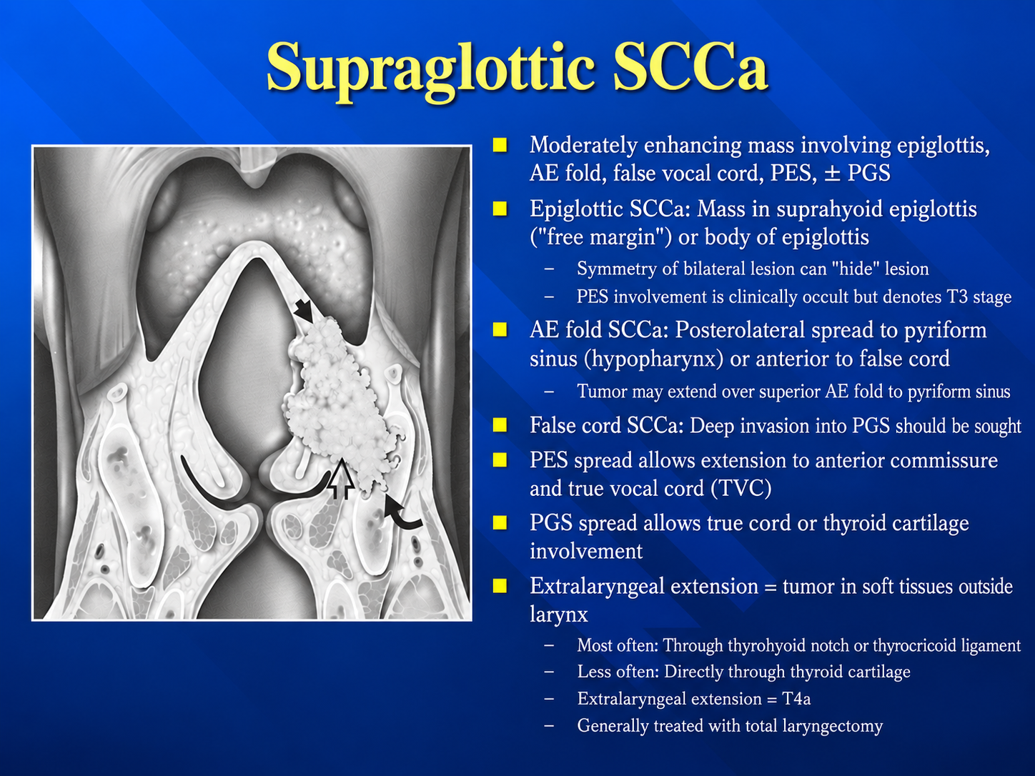
Supraglottic SCC routes of spread
Subsite-Specific Routes of SpreadClick to enlarge
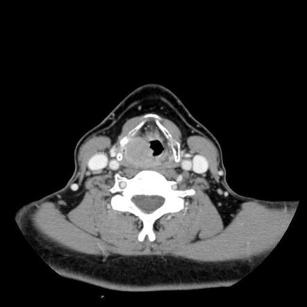
Axial contrast CT example of a supraglottic mass
Axial Contrast CT ExampleClick to enlarge
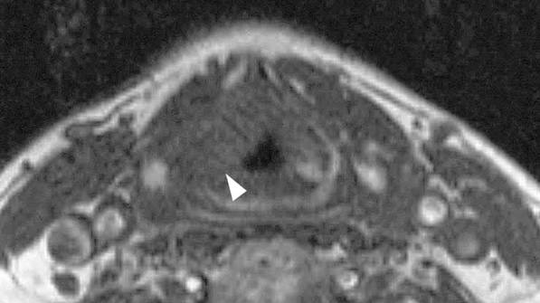
Axial MRI example of a supraglottic lesion
Axial MRI ExampleClick to enlarge

Origin and Behavior

  • Commonly arises from the laryngeal epiglottis, aryepiglottic fold, false cord, arytenoid region, or ventricle.
  • Most invasive tumors are conventional SCC.
  • Symptoms can be delayed because early lesions may not disturb true-cord vibration.
  • Typical imaging appearance is an asymmetric mildly to moderately enhancing mucosal or submucosal mass.
Clinical correlationVocal-cord fixation is determined clinically/endoscopically and should not be inferred from static imaging alone.

Deep-Space Spread

  • Epiglottic primary: preferentially replaces pre-epiglottic fat.
  • False-cord primary: preferentially enters paraglottic fat and may spread transglottically.
  • Aryepiglottic-fold primary: may extend toward the medial piriform sinus, epiglottis, and paraglottic space.
  • Masslike enhancing replacement contiguous with tumor is more convincing than isolated fat stranding.
T3 thresholdPreepiglottic or paraglottic-space invasion makes the primary at least T3.

Nodal Pattern

  • The supraglottis has rich and frequently bilateral lymphatic drainage.
  • First-echelon spread usually involves upper and middle jugular chains, especially levels II and III.
  • Assess both sides for morphology, necrosis, clustering, and extranodal extension—not size alone.
  • Document nodal level, side, number, largest dimension, necrosis, and definite extranodal extension.

Imaging Approach

ModalityBest useLimitation
Contrast CTAirway, cortical cartilage, nodes, rapid stagingNonossified cartilage and dental artifact
MRIDeep spaces, tongue base, nonossified cartilage, equivocal extensionMotion and inflammatory overcall
FDG-PET/CTAdvanced-stage nodal/distant staging and synchronous tumorsInsufficient anatomic resolution for subtle local T staging
Technique: Use thin sections and multiplanar reformations. Sagittal imaging is particularly valuable for the epiglottis and pre-epiglottic space.

Cartilage and Extralaryngeal Spread

  • CT signs include sclerosis, erosion, lysis, and tumor beyond cartilage.
  • Sclerosis is sensitive but nonspecific; destructive change and extra-cartilaginous tumor are more specific.
  • Isolated arytenoid sclerosis should not be equated with invasion.
  • State the deepest confidently involved thyroid-cartilage layer.
  • Assess the thyrohyoid membrane, strap muscles, thyroid gland, deep tongue muscles, hypopharynx, trachea, and neck soft tissues.
Staging distinction: Inner thyroid cortex invasion is T3. Tumor through the outer cortex or extending beyond the larynx is T4a.

AJCC Primary Tumor Categories

CategorySupraglottic definition
TisCarcinoma in situ.
T1One supraglottic subsite with normal vocal-cord mobility.
T2Adjacent supraglottic/glottic mucosa or mucosa outside the supraglottis, without laryngeal fixation.
T3Limited to larynx with vocal-cord fixation and/or postcricoid, pre-epiglottic, paraglottic, or inner thyroid cortex invasion.
T4aThrough outer thyroid cortex and/or tissues beyond the larynx.
T4bPrevertebral space, carotid encasement, or mediastinal invasion.
Current system: AJCC 8th edition remains current for larynx in 2026; Version 9 replaces individual disease sites only as new protocols take effect.

Staging Search Pattern

  1. Define the epicenter and involved supraglottic subsites.
  2. Measure in three dimensions and record midline crossing.
  3. Describe airway narrowing or urgent obstruction.
  4. Map extension to vallecula, tongue base, medial piriform sinus, postcricoid region, glottis, and subglottis.
  5. Assess pre-epiglottic and paraglottic spaces.
  6. State transglottic extension.
  7. Classify thyroid cartilage as normal, equivocal, inner cortex, or through outer cortex.
  8. Assess cricoid and cricoarytenoid joints.
  9. Map extralaryngeal extension.
  10. Evaluate bilateral cervical nodes and distant/synchronous disease when included.

Reporting Checklist

Primary: epicenter, subsites, dimensions, midline, airway.
Deep extent: pre-epiglottic space, paraglottic space, transglottic spread, postcricoid region.
Framework: thyroid inner/outer cortex, cricoid, cricoarytenoid joints.
Beyond larynx: membranes, strap muscles, thyroid, trachea, tongue, esophagus, prevertebral/carotid/mediastinal compartments.
Nodes: level and side, size, necrosis, and definite extranodal extension.

Example Impression

Infiltrative supraglottic mass centered in the left false cord and laryngeal surface of the epiglottis, with replacement of the left paraglottic and pre-epiglottic fat. No definite thyroid or cricoid cartilage invasion and no extralaryngeal extension. Findings indicate at least cT3 disease; final T category should incorporate endoscopic vocal-cord mobility. Bilateral level II–III metastatic adenopathy, including a necrotic left level IIA node with definite imaging extranodal extension.

Important mimics

Consider lymphoma, neuroendocrine or minor-salivary tumors, amyloidosis, inflammatory disease, hematoma, vascular malformation, and laryngocele when the lesion is smooth, submucosal, unusually homogeneous, or not typical for SCC.
Authoritative References

Secondary Links