Epidemiology and Types
CAA may be sporadic, familial, or iatrogenic. Sporadic CAA is most common and may be incidental: GRE screening detects it in up to 16% of asymptomatic older adults. Autopsy prevalence rises from about 5–9% at age 60–69 to 43–58% after age 90.
- Sporadic: age-related impairment of Aβ clearance.
- Familial: rare, usually autosomal dominant, with earlier onset.
- Iatrogenic: rare Aβ seeding after cadaveric dura or pituitary growth hormone, often after a latency of about three decades.
Diagnosis
Boston criteria 2.0 combine clinical, MRI, and pathologic features. Definite diagnosis requires tissue, while probable and possible CAA can be established clinicoradiologically.
- Edinburgh criteria support evaluation of lobar hemorrhage on CT when MRI is unavailable.
- Modified Queen Square criteria have been proposed for iatrogenic CAA.
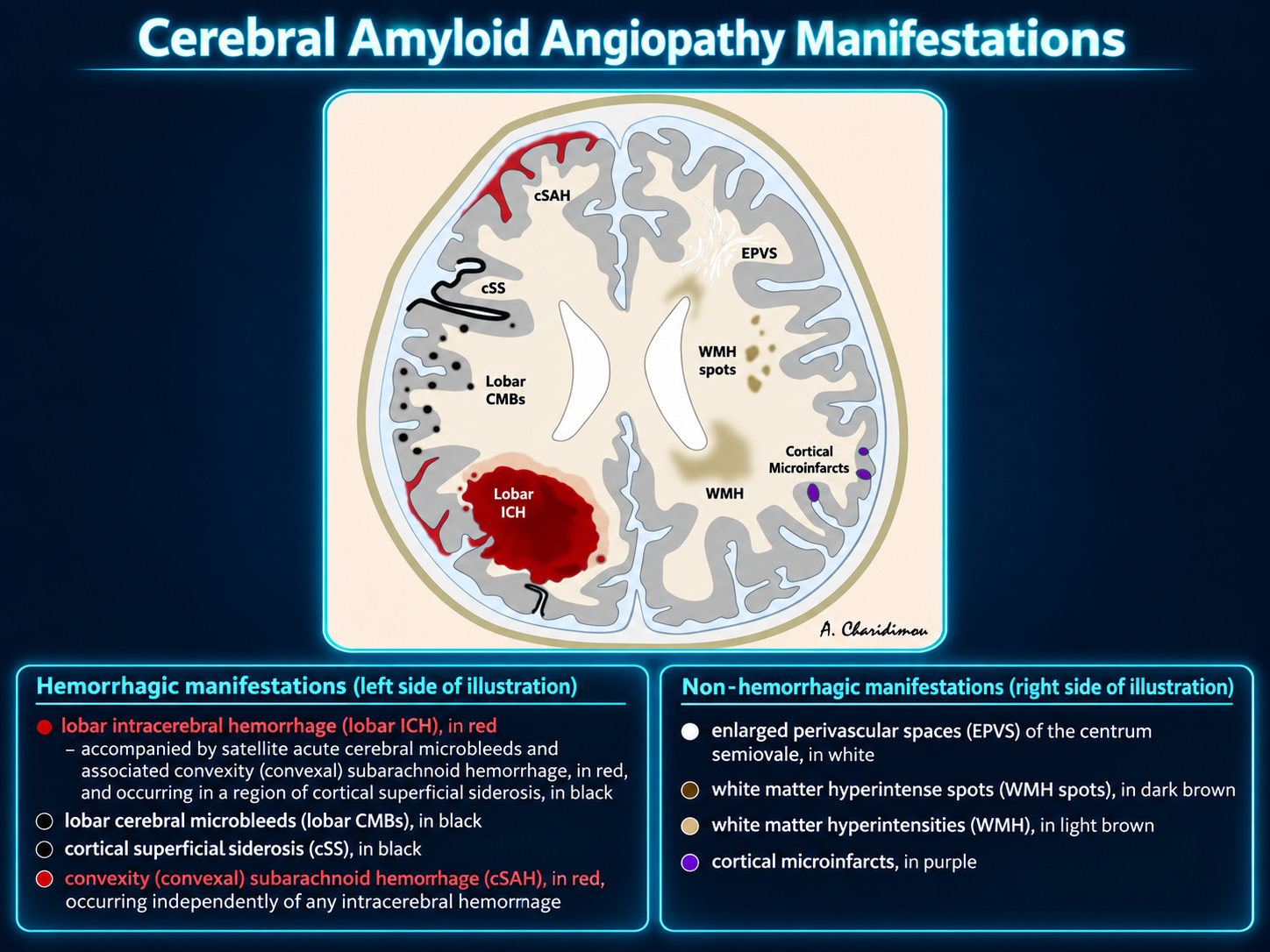
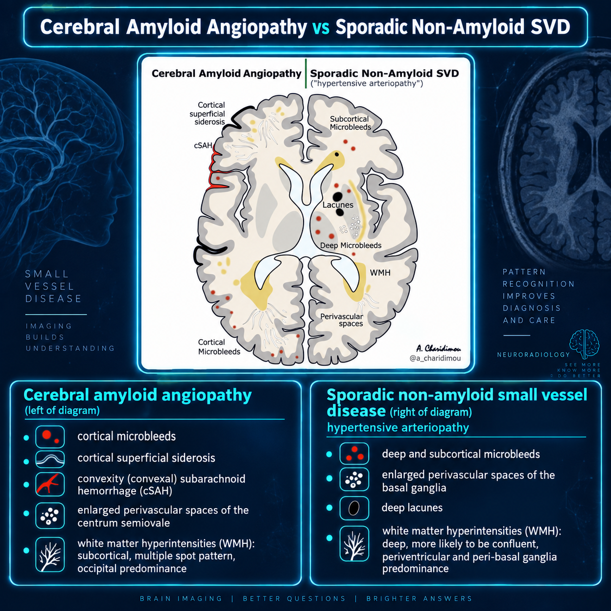
CAA pattern: strictly lobar hemorrhagic lesions, cortical superficial siderosis, convexity SAH, centrum-semiovale perivascular spaces, posterior WMH, and relative sparing of deep nuclei and pons.
Clinical Presentation
- Lobar or superficial cerebellar hemorrhage with focal deficits, headache, seizures, or reduced consciousness.
- Gradual cognitive decline from accumulating microbleeds, ischemic injury, microinfarcts, and lobar lacunes.
- Stepwise decline from recurrent hemorrhage.
- Rapid decline in inflammatory CAA.
- Transient focal neurologic episodes (“amyloid spells”) from convexity SAH, often recurrent and spreading with positive or negative symptoms.
Pathology and Associations
Aβ42 deposition affects small and medium cortical and leptomeningeal arteries, causing fibrinoid degeneration, “double-barreling,” and microaneurysm formation. Congo red produces apple-green birefringence.
- Low CSF Aβ40 and Aβ42.
- CAA pathology occurs in approximately 80–90% of Alzheimer disease.
- About 40% of patients with CAA have Alzheimer disease.
- Associated with Down syndrome and chronic traumatic encephalopathy.
- Usually unrelated to systemic amyloidosis.
Hemorrhagic Imaging Manifestations
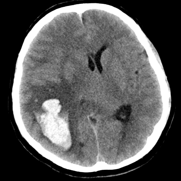
Lobar intracerebral hemorrhage
- Cortico-subcortical, often occipital or temporal; may involve superficial cerebellum.
- Finger-like projections or an island sign may support CAA.
- Basal ganglia and pons are typically spared.
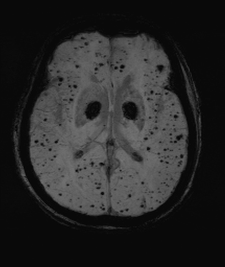
Microbleeds
- 2–10 mm lobar/corticosubcortical susceptibility foci, often occipital-predominant.
- Seen only on T2*-sensitive GRE, EPI, or SWI; not CT or conventional T1/T2/FLAIR.
Other hemorrhage
- Convexity SAH localized to cortical sulci with basal-cistern sparing.
- Cortical or cerebellar superficial siderosis.
- Possible association with intraventricular or atraumatic subdural hemorrhage.
Nonhemorrhagic Imaging
- Posterior/periventricular WMH without U-fiber involvement.
- Cortical microinfarcts (<4 mm) and lobar lacunes (3–15 mm).
- Enlarged perivascular spaces in centrum semiovale and sometimes juxtacortically, with basal-ganglia sparing.
- Cortical atrophy, best assessed with surface reconstruction.
- Rare occipital cortical calcification, especially familial forms.
- Patchy concentric vessel-wall enhancement may occur and may correlate with ischemic risk.
Amyloid PET
PiB, florbetapir, and flutemetamol may show increased cortical tracer uptake with loss of gray-white differentiation.
Distinction from Alzheimer disease is difficult, but an occipital-to-global uptake ratio greater than 1 may favor CAA. Amyloid PET may also help identify coexistence of deep perforator arteriopathy and CAA.
Treatment and Prognosis
No established disease-modifying therapy exists. Investigational approaches include antibody-mediated Aβ removal, reduced production, and increased physiologic clearance; antibody treatment may cause ARIA.
Anticoagulants and thrombolytics may increase disabling hemorrhage risk; evidence-based use requires individualized risk assessment. Indicated antiplatelet therapy may be safe.
Rebleeding risk is greatest in the first three months. Numerous microbleeds, diffuse siderosis, and extensive WMH predict worse outcome.
Differential Diagnosis
- Hypertensive arteriopathy: deep basal-ganglia, pontine, and cerebellar hemorrhages; no convexity SAH or cortical siderosis.
- Multiple cavernomas: random size and distribution; larger characteristic cavernomas may coexist.
- Hemorrhagic metastases: variable size and enhancement.
- Diffuse axonal injury: gray-white junction, corpus callosum, and brainstem in a trauma pattern.
- Fat embolism: appropriate history, walnut-kernel microbleeds, and DWI starfield pattern.
- Radiation vasculopathy: microbleeds conform to the treatment field.
- Post-cardiac surgery change.
- ARIA: may be indistinguishable, but occurs during amyloid-lowering therapy.