Imaging Strategy
| Modality | Best use | Limitation |
|---|
| Contrast CT | Airway, cortical cartilage, nodes, rapid staging | Subtle superficial disease and nonossified cartilage |
| MRI | Paraglottic space, commissures, nonossified cartilage, equivocal extension | Motion and inflammatory overcall |
| FDG-PET/CT | Advanced whole-body staging and post-treatment response | Insufficient resolution for fine mucosal staging |
Technique: Use thin-section contrast imaging with axial planes aligned to the cords and coronal reformations for craniocaudal extension.
Cartilage and Extralaryngeal Spread
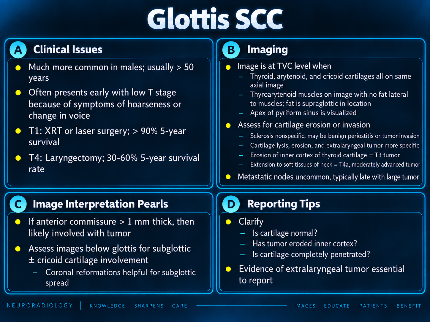
- Do not overcall isolated sclerosis, especially arytenoid sclerosis.
- Confidence increases with focal lysis, cortical breakthrough, matching tumor signal, or an extra-cartilaginous mass.
- Inner thyroid cortex: T3.
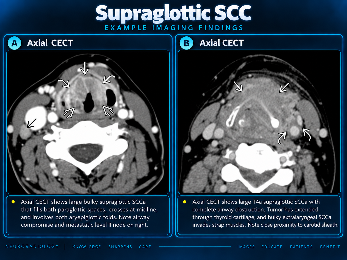
- Outer thyroid cortical penetration: T4a.
- Cricoid invasion: T4a for a glottic primary.
- Beyond larynx: T4a even without a clearly seen cartilage route.
Describe the structure: State strap muscle, thyroid, tracheal, esophageal, deep-tongue, prevertebral, carotid, or mediastinal involvement rather than only saying “extralaryngeal.”
AJCC Primary Tumor Categories
| Category | Glottic definition |
|---|
| Tis | Carcinoma in situ. |
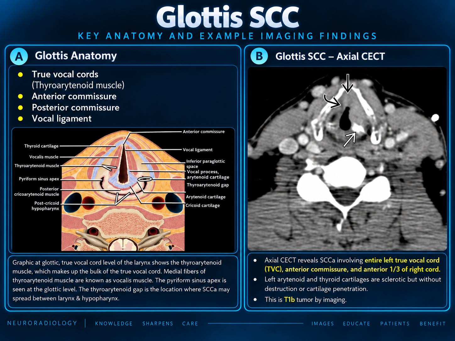
| T1a | One vocal cord; normal mobility. |
| T1b | Both vocal cords; normal mobility. |
| T2 | Supraglottic/subglottic extension and/or impaired mobility. |
| T3 | Limited to larynx with fixation, paraglottic invasion, and/or inner thyroid cortex invasion. |
| T4a | Through outer thyroid cortex, cricoid invasion, and/or tissues beyond the larynx. |
| T4b | Prevertebral space, carotid encasement, or mediastinal invasion. |
Current system: AJCC 8th edition remains current for larynx in 2026. Staging combines radiologic depth with endoscopic mobility.
Level-by-Level Search Pattern
- Confirm the side and anterior/middle/posterior cord epicenter.
- Measure the lesion and characterize superficial versus infiltrative morphology.
- Inspect both commissures and the contralateral cord.
- Evaluate supra- and subglottic extension; measure subglottic length.
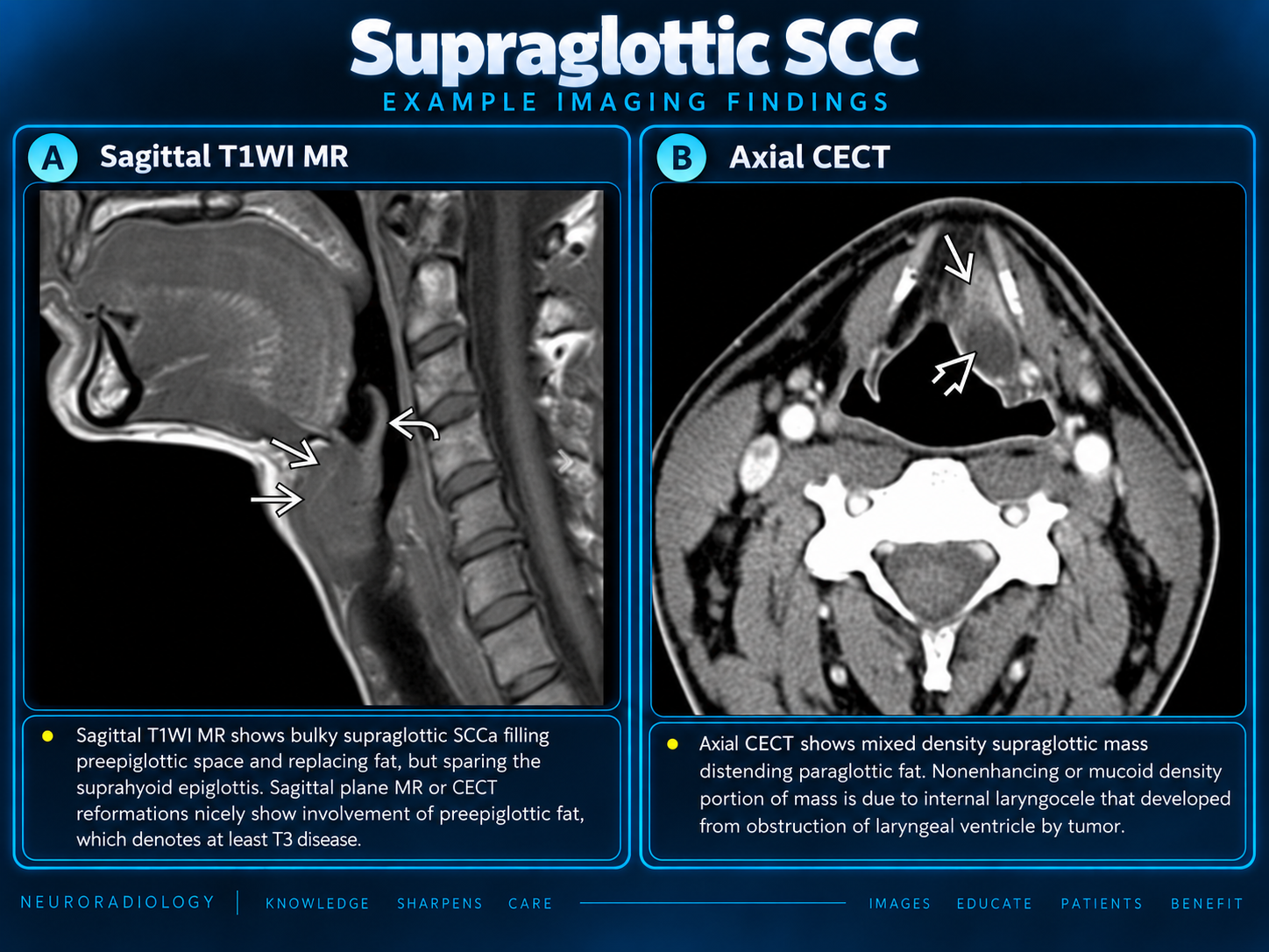
- Assess the paraglottic space and cricoarytenoid units.
- Classify thyroid cartilage as normal, equivocal, inner cortex, or outer breakthrough.
- Inspect the cricoid and cricothyroid membrane.
- Map extralaryngeal spread and airway narrowing.
- Review bilateral levels II–IV and level VI when subglottic extension is advanced.
- Correlate the final T category with endoscopic mobility.
Reporting Checklist
Primary: side, cord segment, dimensions, morphology.
Local extent: anterior/posterior commissure, opposite cord, supra/subglottic spread, paraglottic space.
Framework: cricoarytenoid unit, thyroid cortex, cricoid cartilage.
Beyond larynx: cricothyroid membrane, strap muscles, thyroid, trachea, esophagus, deep tongue.
Nodes: level, side, size, necrosis, and definite extranodal extension.
Example Impressions
At least cT2N0: Enhancing mass centered in the anterior left true vocal cord with anterior-commissure involvement and approximately 7 mm of anterior subglottic extension. No contralateral cord, paraglottic-space, cartilage, or extralaryngeal invasion. Final T category should incorporate endoscopic mobility.
cT3N0: Infiltrative right glottic mass involving the right paraglottic space and inner cortex of the adjacent thyroid cartilage, without outer cortical breakthrough or extralaryngeal extension. Correlate with laryngoscopy for cord fixation.
Selected mimics
Focal laryngitis, polyp, nodule, granuloma, papillomatosis, dysplasia, amyloidosis, cricoarytenoid inflammation, intubation injury, and post-treatment change.