Subglottic Cancer

Core concept: Primary subglottic cancer arises below the true cords and above the lower cricoid border. Because it can grow circumferentially and submucosally before causing symptoms, imaging must define the epicenter, airway caliber, cricoid and thyroid cartilage, tracheal and extralaryngeal extension, and central-compartment nodes through the superior mediastinum.

Visual Reference

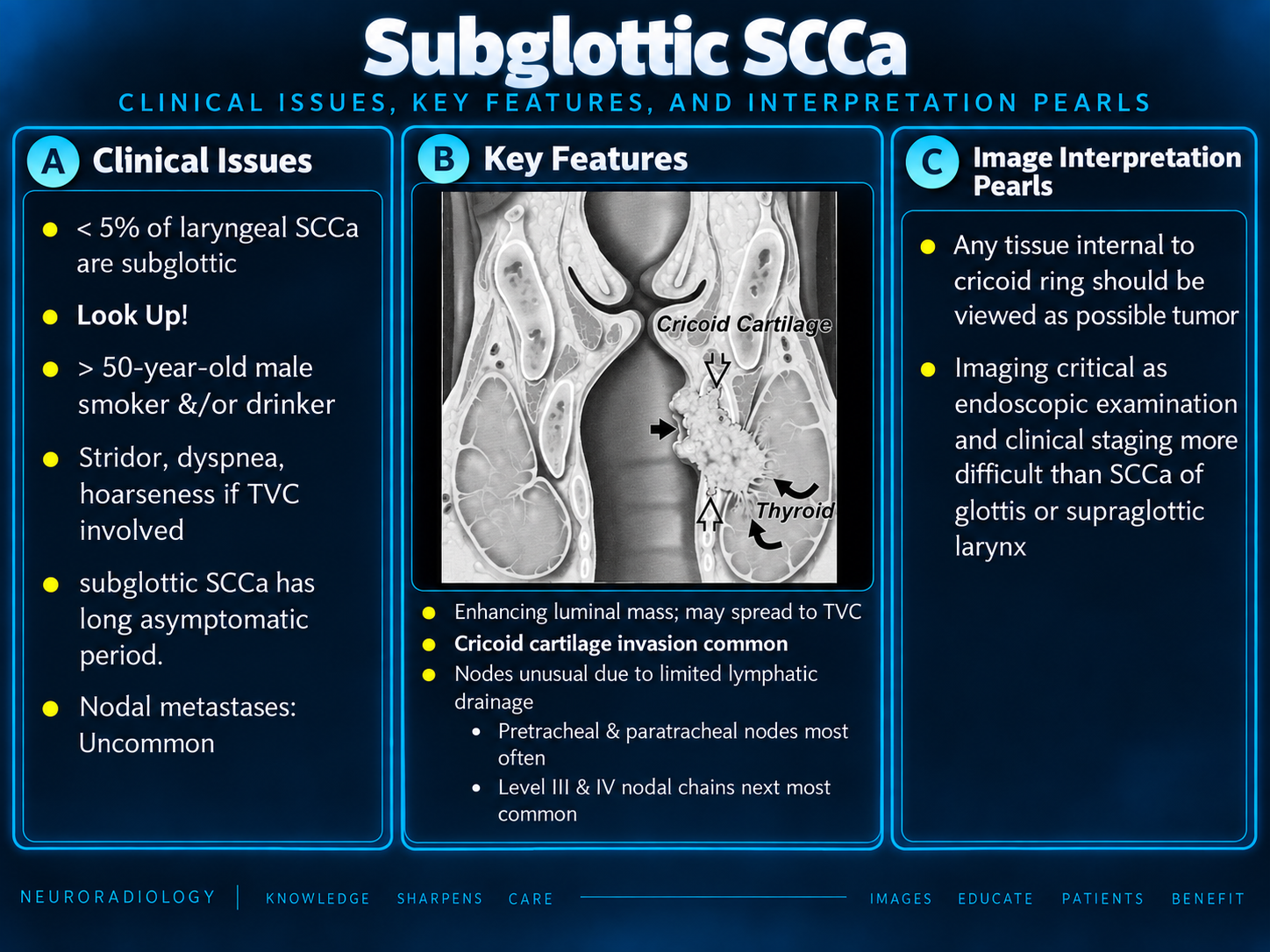
Subglottic SCC clinical issues, key features, and interpretation pearls
Clinical and Imaging OverviewClick to enlarge
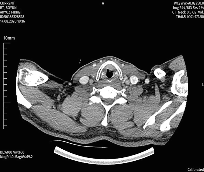
Axial contrast CT example of a subglottic lesion
Axial Contrast CT ExampleClick to enlarge
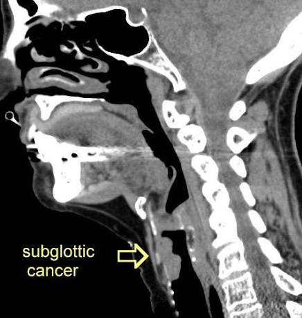
Sagittal CT showing subglottic cancer
Sagittal Airway and Craniocaudal ExtentClick to enlarge

Origin and Presentation

  • Primary subglottic carcinoma is uncommon; most cases are SCC.
  • Confirm that the bulk is below the cords rather than inferior spread from a glottic/transglottic primary.
  • Submucosal growth can delay symptoms.
  • Presentation includes progressive dyspnea, stridor, cough, hemoptysis, and hoarseness after superior extension.
Airway first: Marked narrowing or stridor requires urgent multidisciplinary airway planning before elective imaging or biopsy.

Extent to Measure

  • Three-dimensional size and craniocaudal length.
  • Focal, eccentric, semicircumferential, or circumferential morphology.
  • Percentage of airway circumference and narrowest residual lumen.
  • Distance to and involvement of the true cords.
  • Relationship to the lower cricoid border.
  • Number and length of involved tracheal rings.

Central Nodal Drainage

  • Level VI: Delphian/prelaryngeal, pretracheal, and bilateral paratracheal nodes.
  • Levels III–IV: middle and lower jugular chains.
  • Level VII: superior mediastinal nodes below the suprasternal notch.
  • Report level, side, size, necrosis, and definite extranodal extension.
CoverageExtend imaging through the superior mediastinum; central nodes may be missed if the study stops at the thoracic inlet.

Imaging Strategy

ModalityBest useLimitation
Contrast CTAirway, cortical cartilage, trachea, thyroid, nodes, mediastinumSubmucosal disease and nonossified cartilage
MRICartilage marrow, esophageal interface, perineural spread, edema problem solvingMotion and inflammatory overcall
FDG-PET/CTAdvanced whole-body staging and post-treatment responseLimited detail for exact mucosal boundaries
Technique: Thin-section axial CT with coronal and sagittal reformations through the lower neck and superior mediastinum; review both soft-tissue and bone windows.

Epicenter, Airway, and Spread

  • Primary subglottic origin is favored when the tumor bulk lies below the true cords with superior extension.
  • If no reliable epicenter remains, describe the full extent as transglottic and state the staging limitation.
  • Anterior spread may cross the cricothyroid membrane into strap muscles or thyroid isthmus.
  • Posterior spread may involve posterior cricoid, postcricoid hypopharynx, cervical esophagus, or prevertebral tissues.
  • Lateral spread may involve thyroid lobes, recurrent laryngeal nerve region, and carotid spaces.

Cartilage and Extralaryngeal Disease

  • Describe cricoid as uninvolved, suspicious, or definitely invaded.
  • Assess thyroid and arytenoid cartilage plus both cricoarytenoid joints.
  • Destruction, fragmentation, cortical breakthrough, or adjacent soft-tissue tumor is more specific than sclerosis alone.
  • Evaluate the trachea, thyroid gland, strap muscles, cervical esophagus, prevertebral space, carotid space, and mediastinum.
Subsite-specific rule: Any cricoid or thyroid cartilage invasion in a primary subglottic tumor is T4a.

AJCC Primary Tumor Categories

CategorySubglottic definition
TisCarcinoma in situ.
T1Limited to the subglottis.
T2Extends to vocal cord(s) with normal or impaired mobility.
T3Limited to the larynx with vocal-cord fixation.
T4aInvades cricoid or thyroid cartilage and/or tissues beyond the larynx.
T4bPrevertebral space, carotid encasement, or mediastinal invasion.
Current system: AJCC 8th edition remains current for larynx in 2026. Cord mobility and fixation require endoscopic correlation.

Reporting Checklist

  1. State the epicenter and confidence in primary subglottic origin.
  2. Describe morphology, dimensions, length, circumference, and airway caliber.
  3. Record cord involvement and request endoscopic mobility correlation.
  4. Measure inferior extension and involved tracheal rings.
  5. Assess cricoid, thyroid, arytenoids, and cricoarytenoid joints.
  6. Map cricothyroid membrane and lower-cricoid spread.
  7. Describe thyroid, tracheal, esophageal, strap-muscle, prevertebral, carotid, or mediastinal invasion.
  8. Review Delphian, pretracheal, paratracheal, lower-jugular, and superior-mediastinal nodes.
  9. Report nodal necrosis and definite extranodal extension.
  10. Assess lungs and synchronous aerodigestive disease when included.

Example Impressions and Differential

cT1N0 example: Eccentric enhancing right subglottic mass confined below the true cords and above the lower cricoid border, without cartilage invasion or extralaryngeal extension. No cervical, paratracheal, or superior mediastinal adenopathy.
At least cT4a example: Circumferential subglottic mass centered within the cricoid ring with superior extension to both true cords, inferior extension to the first two tracheal rings, destructive anterior cricoid invasion, and extension through the cricothyroid membrane into strap muscles.

Selected mimics

Adenoid cystic carcinoma, cricoid chondrosarcoma, inflammatory stenosis, granulomatous disease, papillomatosis, post-intubation injury, lymphoma, amyloidosis, and vascular lesions.

Authoritative References

Secondary Links